
A 45-year-old man with a history of smoking presented to the emergency department with complaints of acute chest pain radiating to the left shoulder. Electrocardiogram demonstrated normal sinus rhythm with Q wave in lead III. No ST elevation was seen. Cardiac troponin levels were elevated, suggesting a clinical diagnosis of non-ST segment elevation myocardial infarction.
The patient subsequently underwent a catheter coronary angiography. The left main coronary artery (LMCA) could not be selectively cannulated and, in addition, was not visualized on nonselective contrast injection in the left coronary sinus. Selective angiogram of the right coronary artery (RCA), which was orthotopically arising from the right coronary sinus (RCS), revealed no obstructive disease; however, the left circumflex (LCx) artery was seen arising from the ostio-proximal RCA coursing caudally and then moving cranially towards the obtuse margin of the heart, suggesting anomalous origin of the LCx from the RCA. The left anterior descending (LAD) artery was not seen in this injection (Figure 1).
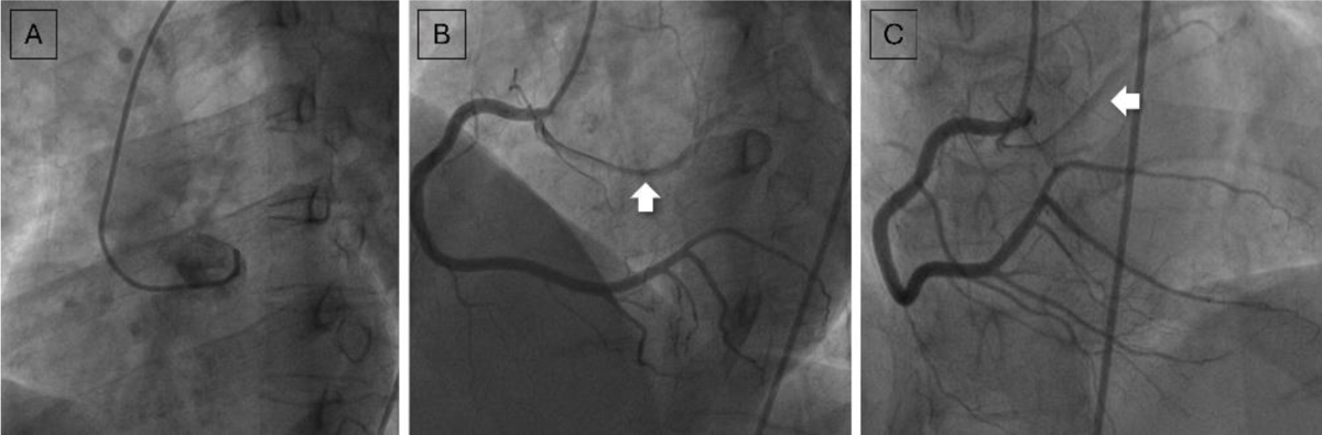
Figure 1
(A) Nonselective contrast injection in the left coronary sinus does not show the left main coronary artery. (B, C) Selective right coronary artery (RCA) angiograms show orthotopically arising RCA and anomalous origin of the left circumflex artery (thick white arrow) from the ostio-proximal RCA. The left anterior descending artery was not seen in this injection.
The patient underwent an electrocardiogram-gated coronary computed tomography angiography (CCTA) to further delineate the anomalous coronary anatomy. CCTA confirmed the anomalous origin of the LCx artery from the proximal RCA, taking a retroaortic course to reach the left atrioventricular groove. Additionally, the LAD artery was seen arising from the RCS, adjacent to the RCA ostium, taking a pre-aortic, interarterial course (between the aorta and main pulmonary artery) to reach the anterior interventricular groove. Thus, a unique circumaortic (retroaortic and preaortic) configuration of the left coronary arteries (LCx and LAD arteries, respectively) was demonstrated on CCTA (Figure 2). No obstructive coronary artery disease or dynamic compression of the LAD artery was seen. The patient was managed conservatively.
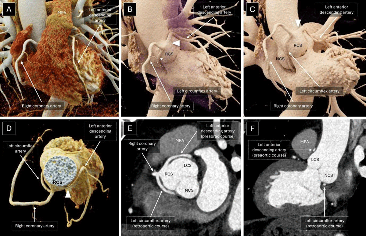
Figure 2
Volume-rendered images (A-D) of coronary computed tomography angiography shows normal origin and course of the RCA. Note the anomalous origin of the LCx from the proximal RCA, taking a retroaortic course to reach the left atrioventricular groove. The LAD artery (white arrowhead) is seen arising from the RCS, adjacent to the RCA ostium, taking a pre-aortic, interarterial course (between the aorta and MPA) to reach the anterior interventricular groove. A short axis reconstruction (E) at the level of the aortic root and an oblique sagittal reconstruction (F) demonstrate the unique circumaortic (retroaortic and preaortic) configuration of the left coronary arteries (LCx and LAD arteries respectively). MPA: main pulmonary artery; LCS: left coronary sinus; NCS: non-coronary sinus; RCA: right coronary artery; LAD: left anterior descending; RCS: right coronary sinus; LCx: left circumflex
Discussion
Coronary artery anomalies can be divided into anomalies of origin, course, or terminations of any of the epicardial coronary arteries. The estimated prevalence of anomalous aortic origin of coronary arteries in adults undergoing coronary imaging is around 2%, with the most common entity being anomalous origin of the RCA from the left coronary sinus, seen in approximately 1.2% cases.1 Of the 1,549 adult patients included in the study, 4 (0.26%) patients had anomalous origin of the LCx artery with retroaortic course, while 1 (0.06%) patient had the LAD artery arising from the RCS with an interarterial course. Coexistence of these two variants as seen in our case is an exceedingly rare anatomical configuration with only a few case reports previously published.2, 3, 4 While an anomalous origin of the LCx artery with a retroaortic course is a benign entity, an anomalous LAD artery arising from the RCS with an interarterial course may predispose to myocardial ischemia and sudden cardiac death.5
Recent 2025 American College of Cardiology/American Heart Association/Heart Rhythm Society/International Society for Adult Congenital Heart Disease/Society for Cardiovascular Angiography & Interventions guidelines recommend exercise testing or dobutamine stress testing with echocardiographic, nuclear, or magnetic resonance imaging to assess for inducible hypoperfusion and regional wall motion abnormalities in asymptomatic patients. Patients who are symptomatic or show ischemic changes in stress testing are recommended for surgical intervention.6 Among asymptomatic patients, surgical intervention is reasonable in cases with high-risk imaging findings, including a slit-like ostium, acute ostial angulation or ostial stenosis, or an intramural course.7 In asymptomatic patients without high-risk imaging features and no signs of inducible ischemia on stress testing, surgical intervention does not demonstrate reduction in risk of sudden cardiac death.8 Surgical intervention options include a coronary artery bypass graft or coronary ostium reimplantation.9 In the presence of this anomaly, conventional catheter angiography is challenging due to technical reasons and may lead to an inadvertent misdiagnosis of significant stenosis or occlusion of the coronary artery.
Conclusion
CCTA provides multiplanar reconstruction and volume rendering capabilities, allowing for accurate delineation of uncommon entities. The present case highlights the importance of CCTA in identifying a rare coronary artery anomaly and providing guidance for future endovascular or surgical procedures.