
A 55-year-old man presented with complaints of claudication in the left upper limb for the past year. The patient was hypertensive, nondiabetic, and a nonsmoker. On examination, the left upper limb arterial pulses were feeble, and there was a blood pressure discrepancy between the right and left upper limbs. The patient was referred to our department for computed tomography angiography of the left upper limb, which revealed non-opacification of the proximal left subclavian artery from its origin until the level of the left thyrocervical trunk. The left subclavian artery from the level of the thyrocervical trunk and the left upper limb arteries showed normal contrast opacification. Notably, the left vertebral artery was seen to originate from the left thyrocervical trunk, with a high entry into the transverse foramen at the C4 level. The dominant right vertebral artery had a normal origin and entered the transverse foramina at the C6 level. The left inferior thyroid artery and the left vertebral artery had a common origin from the thyrocervical trunk (Figures 1, 2).
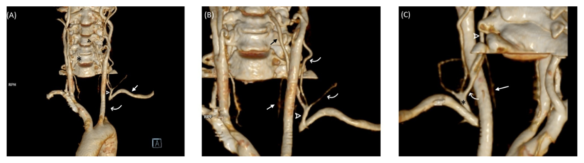
Figure 1
(A) The anterior view of the volume-rendered image shows the left thyrocervical trunk (white arrow) originating from the left subclavian artery (straight arrow). The proximal left subclavian artery was not opacified (curved arrow). The right vertebral artery enters the transverse foramen of C6 (*), while the left vertebral artery is seen to enter the foramen transversarium at C4 level (black arrow). (B) A zoomed image shows the thyrocervical trunk (arrow) giving rise to cervical branches (curved arrows), the left vertebral artery (black straight arrow), and the left inferior thyroid artery (white straight arrow). (C) An oblique posterior view of the volume-rendered image shows a common trunk (curved arrow) of the left inferior thyroid artery (straight arrow) and the left vertebral artery (arrow) arising from the thyrocervical trunk (*).
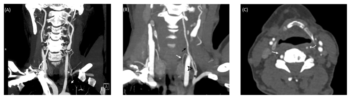
Figure 2
(A) The coronal maximum intensity projection (MIP) image shows non-opacification of the proximal left subclavian artery (arrow) with reformation of the left subclavian artery at the level of the thyrocervical trunk (arrow). The left vertebral artery (curved arrow) is seen originating from the left thyrocervical trunk. (B) The coronal curved reformatted image shows the common trunk (arrow) of the left vertebral artery (black arrow) and the left inferior thyroid artery (white arrow) originating from the thyrocervical trunk (*). (C) An axial image at the level of C6 vertebra shows a dominant right vertebral artery (straight arrow) with an intraforminal course and a smaller left vertebral artery (curved arrow) with an extraforaminal course.
Anatomical variations in the origin of the vertebral artery are common. However, its origin from the thyrocervical trunk has rarely been described. We found two cases where the vertebral artery arose from the thyrocervical trunk.1,2 Another case of rudimentary and accessory left vertebral arteries has been described in the literature, where the accessory left vertebral artery origin was from the left thyrocervical trunk.3 Variations in the vertebral artery origin and course can have surgical and endovascular implications in procedures involving the head and neck.
Competing Interests
The authors have no competing interests to declare.