
Introduction
Pericardial effusions are defined as an abnormal collection of fluid within the pericardial space, exceeding the physiological volume of 15 to 50 mL between the visceral and parietal pericardial layers.1, 2 The clinical significance of pericardial effusions spans a broad spectrum, from an incidental finding on imaging studies performed for other indications to cardiac tamponade, which is a life-threatening condition requiring immediate intervention.3
The pathophysiology of pericardial effusion is closely linked to the pericardium’s unique anatomical and physiological properties. The normal pericardium has limited distensibility, with a steep pressure-volume relationship once its reserve volume is exceeded (Figure 1).4 This fundamental characteristic explains why the rate of fluid accumulation, rather than the absolute volume, primarily determines hemodynamic compromise. Rapidly accumulating effusions may cause tamponade with less than 100 mL of rapidly accumulating fluid, while slowly developing chronic effusions may exceed 1 to 2 liters before producing significant symptoms.5
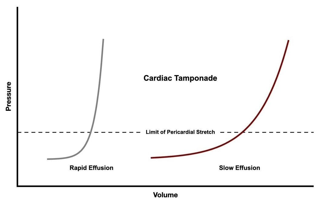
Figure 1
Pressure-volume relationship in pericardial tamponade. Rapid-onset effusions cause tamponades at smaller volumes (grey line) than chronic effusions (red line), allowing gradual pericardial stretch and accommodating larger fluid volumes before hemodynamic compromise.
Cardiac tamponade occurs when intrapericardial pressure rises to levels that impair cardiac filling, resulting in reduced cardiac output and hemodynamic instability.6 The classic Beck’s triad of hypotension, muffled heart sounds, and jugular venous distension, while highly specific, is present in only a minority of patients.7 Pulsus paradoxus, defined as an exaggerated fall in systolic blood pressure of more than 10 mm Hg during inspiration, is a more sensitive clinical finding.8
The advent of multimodality cardiac imaging has revolutionized the diagnosis and management of pericardial diseases. The 2015 European Society of Cardiology (ESC) Guidelines and the 2013 American Society of Echocardiography (ASE) recommendations established echocardiography as the first-line imaging modality for pericardial evaluation.3, 9 More recently, the 2024 Journal of the American College of Cardiology (JACC) Cardiovascular Imaging International Position Statement has further refined the multimodality imaging approach, incorporating advances in cardiac magnetic resonance (CMR) imaging and computed tomography (CT) assessment alongside echocardiographic evaluation.10 The 2025 American College of Cardiology (ACC) Expert Consensus Guidance has provided updated guidance on the comprehensive management of pericardial diseases.11
Epidemiology
Robust epidemiological data on pericardial effusion remain limited, although available evidence suggests it is more prevalent than clinically apparent. Studies from developed countries report an incidence of approximately 3.4% and prevalence ranging from 5.7% to 9% in the general population.12, 13 In emergency department settings, pericardial effusion has been identified in up to 13.6% of patients presenting with dyspnea.14
The etiological distribution of pericardial effusion demonstrates significant geographic variation. In developed countries, idiopathic or presumed viral causes predominate, accounting for approximately 50% of cases, followed by malignancy (10-25%), infectious causes (15-30%), iatrogenic etiologies (15-20%), and connective tissue diseases (5-15%).15, 16 In developing regions, tuberculosis remains the leading cause, representing over 60% of cases, particularly in areas where human immunodeficiency virus coinfection is prevalent.17, 18
Malignancy-associated pericardial effusion carries distinct clinical significance. Autopsy studies demonstrate pericardial involvement in 5% to 15% of patients with malignant neoplasms, with lung cancer (37%), breast cancer (22%), and hematologic malignancies (17%) representing the most common primary tumors.19 Among patients with HIV infection, pericardial effusion has been reported in 5% to 43% depending on inclusion criteria, although the incidence appears to have decreased with widespread use of antiretroviral therapy.20
Post-cardiac surgery pericardial effusion represents an increasingly recognized entity. In a study of 122 consecutive patients undergoing cardiac surgery, effusions were present in 84% by postoperative day 10, though the majority were hemodynamically insignificant.21 Iatrogenic causes, including catheter ablation procedures, percutaneous coronary interventions, and myocardial biopsy, have emerged as essential contributors in contemporary practice, frequently appearing as small rapidly accumulating pericardial effusions with resulting cardiac tamponade.22
Clinical Characteristics
The clinical presentation of pericardial effusion varies widely based on the rate of accumulation, underlying etiology, and presence of associated pericarditis. Patients with slowly accumulating effusions may remain asymptomatic for extended periods, whereas those with rapid accumulation may present with acute hemodynamic compromise.23
Common presenting symptoms include chest discomfort, dyspnea, and a sensation of chest fullness. When pericarditis accompanies effusion, patients typically report pleuritic chest pain that improves with sitting forward and worsens when supine.24
The clinical features of cardiac tamponade reflect impaired cardiac filling and reduced output. The progression typically involves tachycardia as an initial compensatory response, followed by elevated jugular venous pressure, hepatomegaly, and ultimately hypotension and shock.25 Electrical alternans on electrocardiography, characterized by alternating QRS complex amplitudes, represents a highly specific though relatively insensitive finding, occurring in approximately 10% to 20% of cases. Low-voltage complexes are observed more frequently, present in up to 60% of patients with tamponade.26
The ESC has proposed a triage scoring system for patients with cardiac tamponade that integrates etiology, clinical presentation, and imaging findings to guide the urgency of intervention. A score of 6 or greater indicates the need for immediate pericardial drainage, whereas lower scores may permit deferral of drainage for up to 12 to 48 hours under appropriate monitoring.27
Imaging
Role in Diagnosis
Echocardiography
Echocardiography is the primary imaging modality for the detection and assessment of pericardial effusion, endorsed unanimously by major cardiovascular societies.3, 9-11 Its advantages include wide availability, portability for bedside evaluation, absence of ionizing radiation, cost-effectiveness, and the ability to provide real-time hemodynamic assessment.28
On two-dimensional (2D) echocardiography, pericardial effusion appears as an echo-free space between the visceral and parietal pericardium (Figure 2A). The ASE recommends a systematic approach to echocardiographic evaluation that addresses four key components: (1) differentiation between global and localized effusion; (2) quantification of effusion size; (3) description of fluid characteristics; and (4) assessment of hemodynamic impact.9, 29
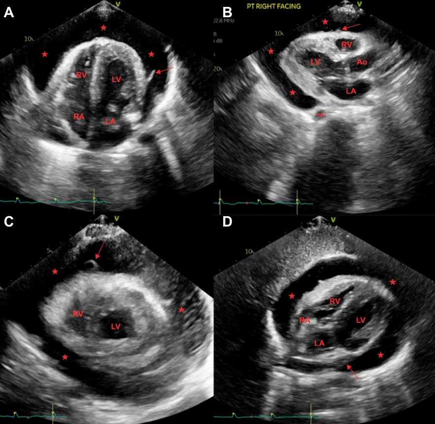
Figure 2
Echocardiographic evaluation of pericardial effusion (standard views). (A) Two-dimensional (2D) apical four-chamber view showing anechoic pericardial space fluid collection (marked as  ) with fibrin strands (marked with arrow) suggestive of pericardial effusion. (B) 2D parasternal long-axis view showing similar fluid collection in the pericardial space (marked as ), notably anterior to the descending aorta, which differentiates from posteriorly located pleural effusion. (C) 2D short-axis view through the mid-left ventricular cavity, and (D) 2D subcostal view showing circumferential pericardial effusion (marked as ). Ao: aorta; DAo: descending aorta; LA: left atrium; LV: left ventricle; RA: right atrium; RV: right ventricle
Effusion sizing is performed by measuring the echo-free space at end-diastole, with the following classification widely adopted: trivial (seen only in systole), small (< 10 mm), moderate (10-20 mm), large (21-25 mm), and very large (> 25 mm).9, 30 These arbitrary criteria were first described in 1984 using M-mode echocardiography and, as a result, poorly describe loculated collections.31 The pericardial effusion distribution should be characterized as circumferential or loculated, with particular attention to posterior and apical collections that may be overlooked in standard views (Figure 2B-D).32 Application of these size classification criteria to cardiac CT and CMR techniques frequently results in discrepancies between sizes called, despite excellent side-by-side correlation between 2D echocardiography and these techniques when similar views are used.
Echocardiographic Signs of Tamponade
Echocardiographic evaluation of cardiac tamponade is a critical competency for practicing cardiologists. The hemodynamic assessment should follow a structured approach evaluating: (1) chamber collapse; (2) respiratory variation in ventricular dimensions; (3) inferior vena cava (IVC) plethora; (4) Doppler flow pattern variations; and (5) hepatic venous flow abnormalities (Figure 3).33, 34
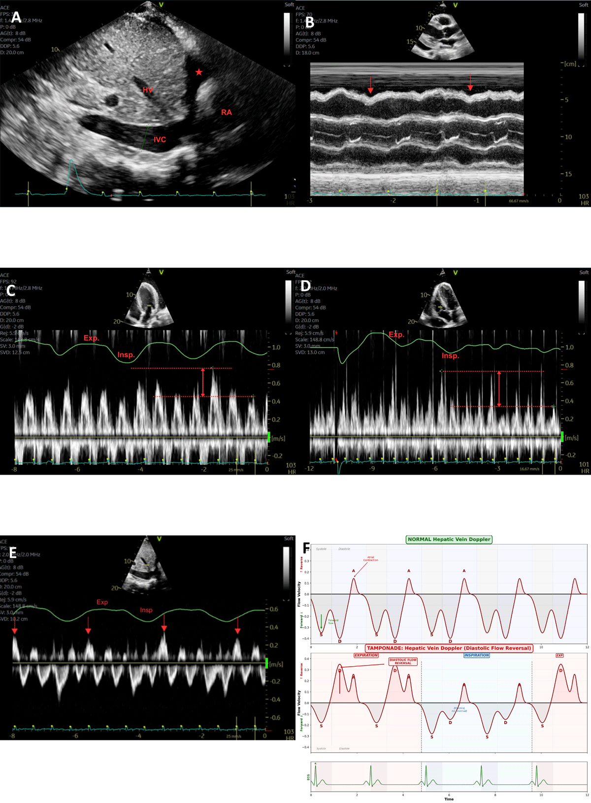
Figure 3
Echocardiographic evaluation of pericardial tamponade. (A) Two-dimensional echocardiography showing typical inferior vena cava plethora (> 20 mm), with blunted respirophasic changes of less than 50%. (B) M-mode echocardiography through RV over one-third of the cardiac cycle in a patient with a large pericardial effusion with tamponade physiology (marked with arrows). (C) Pulse-wave Doppler through the mitral valve inflow shows a variation in peak velocity with respiration in the patient with cardiac tamponade. The peak mitral valve E-velocity of 78 cm/s (expiration) and 43 cm/s (inspiration), with 45% drop in E-velocity. (D) Pulse-wave Doppler through the tricuspid valve inflow in a similar patient showing > 50% drop in velocity with respiration cycle. (E) Pulse-wave Doppler through hepatic veins showing typical flow reversal in cardiac tamponade. (F) Schematic diagram showing comparison of normal biphasic hepatic vein flow on pulse-wave Doppler with diastolic flow reversal in cardiac tamponade. IVC: inferior vena cava; HV: hepatic vein; RA: right atrium; Exp: expiration; Insp: inspiration
Right atrial collapse during ventricular systole represents an early and sensitive sign of elevated intrapericardial pressure. The sensitivity increases when the collapse duration exceeds one-third of the cardiac cycle.35 Right ventricular diastolic collapse occurs subsequently as intrapericardial pressure rises, demonstrating high specificity for hemodynamically significant tamponade (Figure 3B).36 Left-sided chamber collapse, though less common, may occur in cases of loculated effusions, pulmonary hypertension, or elevated right heart filling pressures.37
IVC assessment provides crucial information regarding right atrial pressure. In tamponade, the IVC is typically dilated (> 21 mm) with diminished respiratory variation (< 50% collapse with inspiration or sniff), reflecting elevated and fixed right atrial pressure (Figure 3A).38 While sensitive, IVC plethora is nonspecific and may be observed in other conditions, including volume overload, pulmonary hypertension, and positive-pressure ventilation.39
Doppler assessment of respiratory variation in transvalvular flows constitutes the echocardiographic equivalent of pulsus paradoxus, reflecting enhanced ventricular interdependence in the setting of tamponade physiology. Normal respiratory variation in mitral inflow velocity is < 30%, while tricuspid inflow varies < 60%.40 In tamponade, these thresholds are exceeded, with inspiratory decreases in mitral E-wave velocity > 25% to 30% (Figure 3C) and inspiratory increases in tricuspid E-wave velocity > 40% to 60% (Figure 3D) being diagnostic.41, 42
Hepatic vein Doppler demonstrates characteristic abnormalities in tamponade, with expiratory diastolic flow reversal indicating impaired venous return during the phase when intrathoracic pressure is highest (Figure 3E,F). This finding demonstrates high positive and negative predictive values (82% and 88%, respectively) but requires care in image acquisition and interpretation.43, 44
The “swinging heart” sign, in which the heart oscillates within a large effusion, suggests significant fluid accumulation but does not necessarily correlate with hemodynamic compromise.45 Interventricular septal shift toward the left ventricle during inspiration (“septal bounce”) reflects enhanced ventricular interdependence and supports the diagnosis of tamponade physiology.46
Cardiac Computed Tomography
Cardiac CT provides complementary information to echocardiography, offering a larger field of view, excellent spatial resolution, and the ability to characterize pericardial fluid density by Hounsfield unit (HU) measurements.47 CT is particularly valuable when echocardiographic windows are suboptimal, when loculated effusions are suspected, and for preoperative planning.48
Pericardial fluid characterization by CT can suggest the underlying etiology. Simple transudative effusions typically measure 0 to 20 HU (Figure 4A), while higher attenuation values suggest exudative, hemorrhagic, or purulent content.49 Hemopericardium, as may occur with aortic dissection or myocardial rupture, demonstrates high attenuation (40-60 HU or higher). CT findings suggesting tamponade include flattening or inversion of the right atrial and ventricular walls, distension of the IVC and hepatic veins, and reflux of contrast into the IVC and azygos system (Figure 4B).50, 51
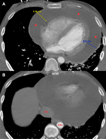
Figure 4
Computed tomography (CT) assessment of pericardial effusion. (A) Contrast-enhanced CT (axial view) demonstrating a large circumferential pericardial effusion (marked as ) with simple fluid attenuation (0-20 Hounsfield units) suggestive of transudative process. (B) Similar patient’s CT showing dilated inferior vena cava, suggesting elevated right atrial pressure.
CT is the imaging modality of choice for detecting pericardial calcification, which may be missed by other modalities. This capability is critical in evaluating constrictive pericarditis and effusive-constrictive disease.52
Cardiac Magnetic Resonance Imaging
CMR provides a comprehensive assessment of pericardial anatomy and physiology without ionizing radiation. Its tissue characterization capabilities allow differentiation between simple and complex effusions based on T1 and T2 signal characteristics.53 Simple effusions appear bright on steady-state free precession cine sequences, with signal intensity similar to or greater than epicardial fat (Figure 5A,D).54
CMR is particularly valuable for detecting pericardial inflammation using late gadolinium enhancement (Figure 5B) and T2-weighted short-tau inversion recovery sequences (Figure 5C).55 Pericardial enhancement on LGE imaging indicates active inflammation and has prognostic value for predicting recurrence and guiding therapeutic decisions, including the use of anti-inflammatory agents and interleukin-1 (IL-1) blockers.56
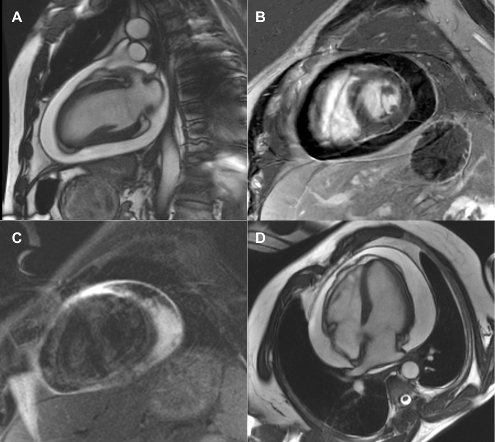
Figure 5
Cardiac magnetic resonance (CMR) assessment of pericardial effusion. (A) A cine still frame of a two-chamber view, and (D) four-chamber view shows a large circumferential pericardial effusion of simple fluid. (B) Late gadolinium enhancement imaging on CMR shows a large circumferential pericardial effusion with mild pericardial enhancement, suggestive of fibrosis. (C) T2-weighted imaging shows increased signal in the visceral and parietal pericardium in suggestive of active pericardial edema.
Real-time cine CMR can demonstrate chamber collapse and respiratory variation in ventricular filling, similar to echocardiography.57 Phase-contrast sequences allow quantification of mitral and tricuspid inflow variation, with cutoffs of ≥ 25% decrease and ≥ 40% increase in inspiratory velocities, respectively, being suggestive of tamponade physiology.58 However, the limited availability and logistical constraints of CMR restrict its utilization. CMR requires prolonged imaging times within a confined scanning magnet, making it unsuitable for evaluation in unstable patients where cardiac tamponade is suspected.
Role of Multimodality Imaging in Differentiation of Pericardial Effusion Mimickers
The 2024 JACC Cardiovascular Imaging International Position Statement by Klein et al. highlights several important mimickers of pericardial effusion that warrant systematic evaluation with multimodality imaging.10 Typical mimickers include:
Epicardial fat—appears as a heterogeneous echodensity that moves synchronously with the myocardium, most commonly anterior to the right ventricle. When echocardiographic findings are equivocal, CT reliably identifies epicardial fat based on its characteristic negative Hounsfield unit attenuation (typically -50 to -100 HU), and CMR demonstrates fat signal on T1-weighted sequences.
Pleural effusion—on transthoracic echocardiogram (TTE), a left pleural effusion may closely mimic a posterior pericardial effusion. The parasternal long-axis view is key: Pericardial effusion lies anterior to the descending thoracic aorta, while pleural effusion tracks posterior to it (Figure 2B). CT provides definitive differentiation with its wider field of view and multiplanar capability.
Pericardial cyst—most located adjacent to the right atrium, pericardial cysts may appear as loculated echo-free spaces on TTE, potentially being mistaken for loculated pericardial effusion. CT demonstrates a well-circumscribed, non-enhancing, homogeneous, water-attenuation structure, while CMR shows a characteristic high T2 signal and no enhancement on LGE sequences.
Pericardial and mediastinal masses—including benign tumors (eg, hemangiomas) and malignant lesions, these may present as heterogeneous echodensities adjacent to the heart on TTE, mimicking complex effusions. CT angiography and CMR provide tissue characterization, essential for differentiation.
Multimodality imaging (TTE, CT, CMR) is essential for confirming pericardial effusion and excluding mimickers when echocardiographic findings are ambiguous, with CT providing spatial/density characterization and CMR offering superior tissue characterization.
Role in Management
Echocardiography
Echocardiography-guided pericardiocentesis represents the contemporary standard of care for percutaneous drainage of pericardial effusion, having largely replaced blind and fluoroscopy-guided approaches.59 The Mayo Clinic experience, encompassing over 1,127 therapeutic procedures over 21 years, established the safety and efficacy of this approach, with procedural success rates of 97% and major complication rates of only 1.2%.60
The echocardiographic guidance of pericardiocentesis involves critical steps. First, a comprehensive echocardiographic examination identifies the largest fluid accumulation and the optimal entry site, which is the point where the effusion is closest to the transducer with no intervening vital structures.61 The Mayo Clinic approach advocates for the selection of the entry site based purely on echocardiographic findings rather than adherence to a single anatomical approach.62
Multiple approaches have been described, including subcostal (subxiphoid), apical, and parasternal routes. Contemporary data suggest that echocardiographically selected sites, most often apical or para-apical, yield higher success rates and lower complication rates than the traditional subcostal approach.63 In a registry of 253 procedures, the apical approach was used in 92% of cases, with an overall success rate of 97% and a complication rate of 3%.64
Echocardiography may be used to define the optimal position and needle trajectory without direct visualization during needle insertion, or, alternatively, an ultrasound needle guide may be used when direct intraprocedural visualization is desired.65 Injection of agitated saline through the needle prior to guidewire insertion and dilatation is essential to confirm intrapericardial positioning, producing a characteristic contrast effect within the pericardial space. A prospective multicenter study demonstrated that real-time echo-monitored pericardiocentesis achieved a 99% success rate with major complications in only 1.2% of cases.66, 67
Extended catheter drainage following initial pericardiocentesis has been shown to reduce effusion recurrence. The Mayo Clinic data demonstrated that prolonged drainage reduced recurrence rates for early and late postoperative effusions by 46% and 50%, respectively. The use of pericardial catheters increased from 23% to 75% over the study period, accompanied by significant reductions in recurrence and the need for surgical intervention.68
Other Multimodality Imaging
The 2024 JACC International Position Statement provides a comprehensive algorithm integrating multimodality imaging in the management of pericardial effusion.10 The approach emphasizes echocardiography as the first-line modality, with CMR and CT reserved for specific clinical scenarios requiring additional anatomical or tissue characterization information.
For patients presenting with pericardial effusion, the initial echocardiographic evaluation should assess effusion size, distribution, and hemodynamic impact. Small effusions without hemodynamic compromise in the setting of acute pericarditis may be managed conservatively with anti-inflammatory therapy and serial imaging.69 Large effusions without tamponade warrant consideration of diagnostic or therapeutic pericardiocentesis, particularly when etiology is unclear.70
CMR is recommended as a second-line test when: (1) echocardiographic windows are suboptimal; (2) there is clinical suspicion of myopericarditis; (3) assessment of pericardial inflammation is needed to guide anti-inflammatory therapy; or (4) characterization of complex effusions or masses is necessary.71, 72 The presence of pericardial LGE may identify patients with transient constrictive pericarditis who may respond to medical therapy rather than requiring pericardiectomy.
CT is preferred when: (1) pericardial calcification is suspected; (2) rapid assessment is needed in hemodynamically unstable patients with poor echocardiographic windows; (3) CT-guided pericardiocentesis is planned for loculated effusions; or (4) evaluation of extracardiac pathology (mediastinal masses, lung disease) is required.73 CT angiography may be essential in cases of suspected aortic dissection or rupture presenting with hemopericardium.74
The integration of imaging findings with clinical presentation and biomarkers optimizes management decisions. Elevated inflammatory markers (C-reactive protein, erythrocyte sedimentation rate) in conjunction with pericardial LGE on CMR support ongoing inflammation and guide the intensity and duration of anti-inflammatory therapy. The emerging role of IL-1 blockers in recurrent pericarditis has been informed by imaging assessment of disease activity.75, 76, 77
Prognosis and Follow-up
The prognosis of pericardial effusion is primarily determined by the underlying etiology. A meta-analysis of 23 studies encompassing 17,022 patients demonstrated that pericardial effusion is associated with a 59% increased risk of mortality (HR 1.59; 95% CI, 1.37-1.85) compared to patients without effusion.78 This prognostic impact was consistent across various underlying conditions, including pulmonary arterial hypertension, chronic heart failure, myocardial infarction, and malignancy.
Malignant pericardial effusion carries a particularly poor prognosis, with a median survival of 2 to 3 months after diagnosis. Studies report 1-year survival rates of 13% to 27% and 2-year survival rates of 9% to 17% following surgical drainage.79, 80 Positive cytology in pericardial fluid and evidence of pericardial metastasis on preoperative imaging are independent predictors of reduced survival. Recurrence rates for malignant effusions range from 24% to 31%, with most recurrences occurring within the first year.81
In contrast, idiopathic pericardial effusion carries a favorable prognosis. Long-term follow-up of patients with clinically significant idiopathic effusions requiring pericardiocentesis demonstrates survival comparable to the general population.82 Pericardiectomy is required in approximately 8% of cases due to recurrence, effusive-constrictive disease, or chronic relapsing pericarditis.83
Follow-up imaging recommendations depend on etiology, effusion size, and clinical trajectory. For patients with small effusions in the context of acute pericarditis, echocardiographic reassessment is recommended after completion of anti-inflammatory therapy to document resolution. Large effusions managed conservatively should undergo serial echocardiography at intervals determined by clinical status, typically weekly until stability is established.84
Following pericardiocentesis, imaging surveillance can monitor for recurrence and development of constrictive physiology. The emergence of echocardiographic features suggesting constriction after drainage of a pericardial effusion should prompt consideration of effusive-constrictive pericarditis, a condition characterized by persistent constrictive hemodynamics despite fluid removal. CMR can help differentiate transient from chronic constrictive disease by identifying persistent pericardial enhancement, suggesting ongoing inflammation amenable to medical therapy.85, 86
Pericardial Effusion in Pulmonary Hypertension
Pericardial effusion occurs in 20% to 26% of patients with pulmonary arterial hypertension and independently predicts mortality, with median survival decreasing from 76.5 months without effusion to 11.3 months with moderate or greater effusion.87 The pathophysiology differs from that of inflammatory or malignant causes: elevated pulmonary vascular resistance increases right ventricular afterload and systemic venous congestion, raising coronary venous pressures and promoting myocardial interstitial fluid formation that transudates across the epicardium into the pericardial space. Echocardiographic diagnosis is challenging because classic tamponade signs (right atrial/ventricular diastolic collapse, IVC plethora, respiratory Doppler variation) may be atypical or absent due to chronically elevated right-sided pressures, chamber hypertrophy, and baseline right heart failure, making left-sided chamber collapse potentially more prominent.88 Management is controversial: small effusions warrant medical optimization with pulmonary hypertension-specific therapy and diuretics, while pericardiocentesis carries substantial risk (40-50% mortality in one case series) because relieving pericardial pressure may cause acute right ventricular dilation, septal flattening, left ventricular compression, and hemodynamic collapse; if necessary, serial low-volume drainage under invasive pulmonary artery catheter monitoring is preferred.89 Multimodality imaging is essential for risk stratification, with echocardiography assessing right ventricular function, right atrial pressure, and serial effusion size; CMR identifying myocardial edema and biventricular function; and CT aiding surgical planning for pericardial window procedures.
Conclusions
Pericardial effusion and cardiac tamponade represent clinical entities that demand proficiency in multimodality cardiac imaging for optimal patient outcomes. Echocardiography maintains its position as the cornerstone of evaluation, providing rapid, noninvasive assessment of effusion characteristics and hemodynamic impact while enabling safe percutaneous intervention. The structured echocardiographic approach—encompassing 2D imaging, M-mode, and Doppler assessment—allows comprehensive evaluation of tamponade physiology at the bedside.
CMR and CT serve as valuable complementary modalities, each with distinct strengths. CMR’s tissue characterization capabilities have transformed the approach to inflammatory pericardial disease, enabling identification of patients who may benefit from intensified medical therapy versus those requiring surgical intervention. CT provides rapid assessment and superior detection of calcification, with utility in emergent settings and for procedural guidance. Special consideration must be given to pericardial effusion evaluation in patients with pulmonary hypertension, where diagnostic criteria may be atypical, and management decisions carry unique hemodynamic risks.
The contemporary evidence base, synthesized in recent position statements from major cardiovascular societies, provides clear algorithms for integrating these modalities into clinical practice. Cardiologists and multimodality imaging specialists must maintain expertise across these imaging platforms to deliver optimal care to patients with pericardial disease. As therapeutic options continue to evolve, particularly with the introduction of IL-1 blockers for recurrent pericarditis, imaging-guided management will become increasingly central to achieving favorable outcomes.
Key Points
Pericardial effusion is a common clinical entity with etiologies varying by geographic region; viral/idiopathic causes predominate in developed countries, while tuberculosis remains the leading cause in developing regions.
The rate of fluid accumulation, rather than absolute volume, primarily determines hemodynamic compromise; acute effusions may cause tamponade with less than 100 mL of fluid accumulation.
Echocardiography is the first-line imaging modality, providing comprehensive assessment of effusion size, distribution, fluid characteristics, and hemodynamic impact.
Key echocardiographic signs of tamponade include right atrial and ventricular collapse, inferior vena cava plethora, and exaggerated respiratory variation in transvalvular Doppler flows (mitral E-wave variation > 25-30%, tricuspid E-wave variation > 40-60%).
Echocardiography-guided pericardiocentesis achieves success rates of 97% to 99% with major complication rates of 1% to 2%, representing a significant improvement over blind technique.
Cardiac magnetic resonance provides superior tissue characterization, with pericardial late gadolinium enhancement indicating active inflammation and guiding decisions regarding anti-inflammatory therapy and IL-1 blockers. Computed tomography is the modality of choice for detecting pericardial calcification and characterizing effusion density through Hounsfield unit measurements.
Extended catheter drainage following pericardiocentesis reduces recurrence rates by approximately 50%.
Malignant pericardial effusion carries a poor prognosis, with median survival of 2 to 3 months; positive fluid cytology and pericardial metastasis on imaging predict worse outcomes.
Pericardial effusion mimickers—including epicardial fat, pleural effusion, pericardial cysts, and mediastinal masses—require systematic evaluation with multimodality imaging for accurate differentiation.
Pericardial effusion in pulmonary arterial hypertension is an independent predictor of mortality; echocardiographic evaluation may show atypical tamponade features, and pericardiocentesis carries unique risks due to potential acute right ventricular dilation and hemodynamic collapse.
Competing Interests
Dr. Luis is a consultant for Kiniksa Pharmaceuticals, Cardiol Therapeutics, and Ventyx Biosciences. The other authors have no competing interests to declare.