
Figure 1
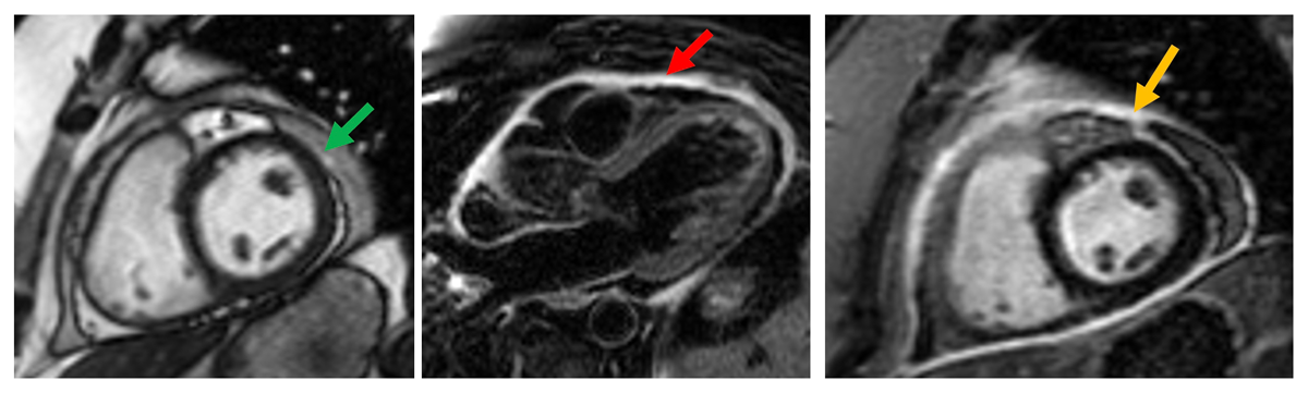
Case example of a 32-year-old man with acute pericarditis. Cine-CMR (left panel) shows a pericardial effusion laterally with heterogenous signal suggesting an exudative effusion. The middle panel shows T2-weighted imaging with fat suppression showing hyperintensity of the pericardium (red arrow). The right-sided panel shows LGE imaging demonstrating circumferential pericardial LGE (orange arrow). CMR: cardiac magnetic imaging; LGE: late gadolinium enhancement
Video 1
Real-time free breathing short-axis cine cardiac magnetic resonance shows respirophasic motion of the interventricular septum and enhanced ventricular interdependence. Note the shift of the septum towards the left ventricle during inspiration; see also at https://vimeo.com/1161598636/ca19594af5.
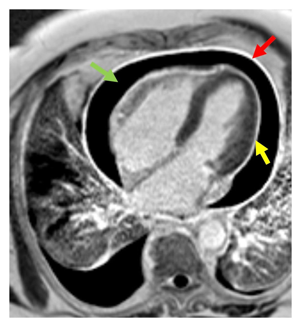
Figure 2
Late gadolinium enhancement (LGE) cardiac magnetic resonance image of a patient with acute pericarditis (phase sensitive inversion recovery sequence). Note the moderate circumferential pericardial effusion (green arrow, jet black pericardial space), and circumferential pericardial LGE involving both the visceral (yellow arrow) and parietal pericardium (red arrow).
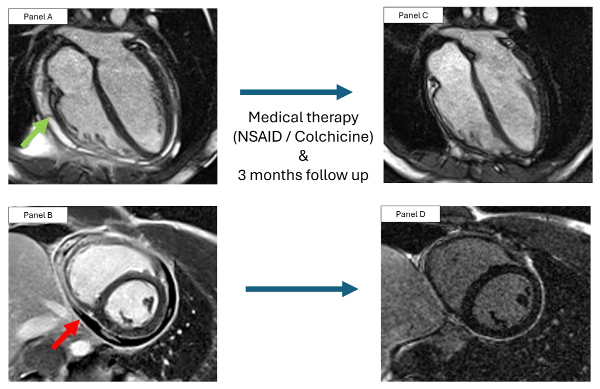
Figure 3
Case example of a 38-year-old man with acute pericarditis. Still image of cine cardiac magnetic resonance (CMR) shows a small circumferential pericardial effusion with adhesions (green arrow, panel A), thickened pericardium, and pericardial late gadolinium enhancement (LGE) (panel B). After medical therapy with nonsteroidal anti-inflammatory drugs and colchicine, follow up CMR 3 months later shows resolution of pericardial thickening and the pericardial effusion (panel C), with mild residual pericardial LGE (panel D).
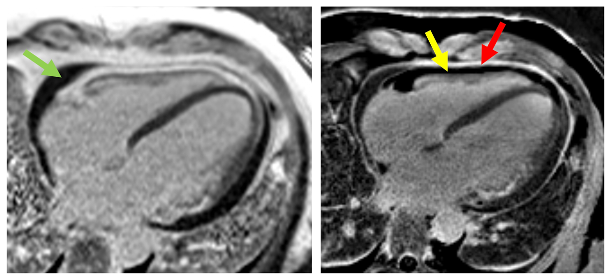
Figure 4
Case example of a 45-year-old patient with pericarditis undergoing standard late gadolinium enhancement (LGE) with phase sensitivity inversion recovery (left panel) and double spectral attenuated inversion recovery (DSPAIR, right panel). The patient has a small pericardial effusion (green arrow) and high signal intensity on the epicardial and the pericardial surface, increasing the difficulty of determining presence of pericardial LGE versus pericardial fat or both. Use of DSPAIR suppresses epicardial fat on the anterior right ventricular wall (yellow arrow) and persistent high signal intensity of the parietal pericardium is present (red arrow) confirming active pericardial inflammation.
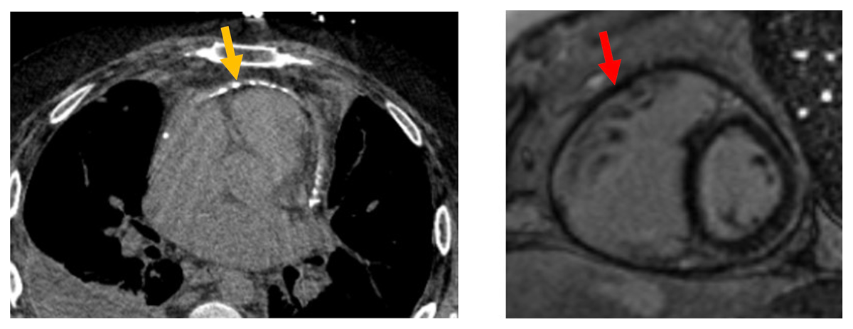
Figure 5
Example case of a patient with advanced end-stage calcific constrictive pericarditis. Note the pericardial calcifications on computed tomography (left panel, orange arrow), pericardial thickening, and absence of late gadolinium enhancement (LGE) on cardiac magnetic resonance (CMR, right panel, red arrow). Care should be applied when interpreting CMR as the lack of pericardial LGE does not exclude a pericardial pathology.
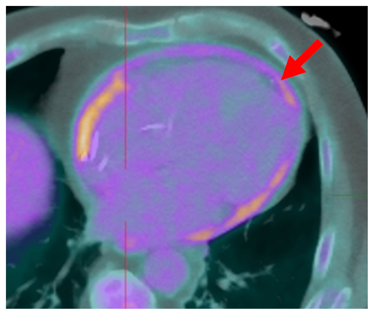
Figure 6
18F-fluorodeoxyglucose positron emission tomography shows circumferential uptake in the pericardium in a patient with established lupus pericarditis.