
A 20-year-old male patient presented to the cardiology outpatient clinic with complaints of intermittent episodes of mild atypical chest pain for the past 3 months. Transthoracic echocardiography (ECG) revealed ectasia and tortuosity of the right coronary artery (RCA) with possible communication with the coronary sinus (CS). An ECG-gated coronary computed tomography (CT) angiography was performed for further anatomical delineation, which revealed a dilated (maximum diameter of 13 mm) and tortuous RCA along its entire course. Adjacent to the crux of heart, a fistulous connection was seen between the distal RCA and the CS with presence of dystrophic calcification along the fistula. There was stenosis of the CS ostium with aneurysmal dilatation of the CS measuring 12.4 × 8.1 × 6.7 cm; no intraluminal thrombus was noted. The dilated CS was seen displacing the heart cranially (Figure 1, Videos 1 and 2).
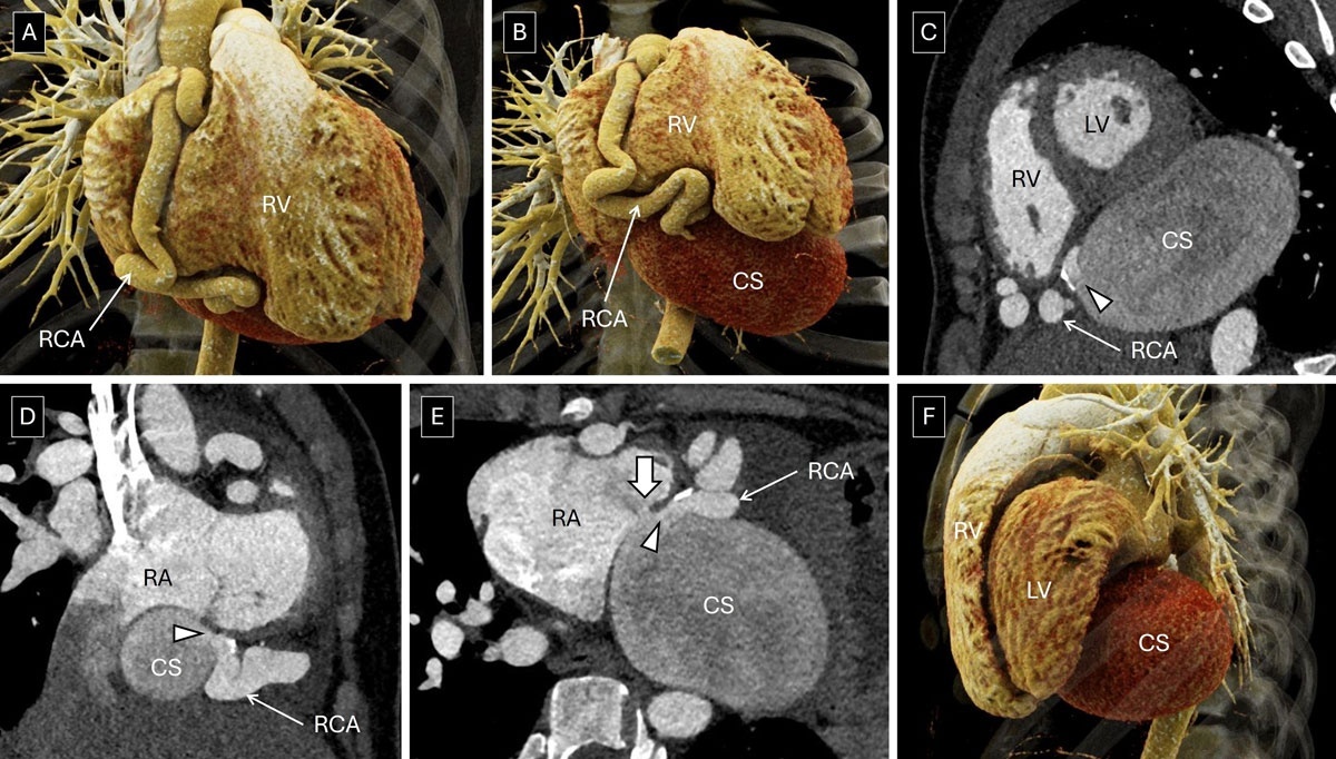
Figure 1
(A, B) Volume-rendered images demonstrate a dilated and tortuous right coronary artery (RCA) along its entire course. (C–E) Multiplanar reconstructions of computed tomography angiography depict a fistulous connection (arrowhead) between the distal RCA and the coronary sinus (CS). Also present is stenosis of the CS ostium (thick white arrow in E) with aneurysmal dilatation of the CS. (F) Volume-rendered image shows the dilated CS displacing the heart cranially. LV: left ventricle; RA: right atrium; RV: right ventricle
Video 1
Volume rendering of computed tomography angiography dataset; see also at https://vimeo.com/1153740220.
Video 2
Volume rendering of computed tomography angiography dataset demonstrating the aorta, right coronary artery, and coronary sinus with the rest of the cardiovascular structures subtracted; see also at https://vimeo.com/1153741063.
Coronary artery fistulas represent abnormal connections between the coronary arterial system and cardiac chambers or other vascular structures; drainage into the CS is seen in 7% to 9% of cases, resulting in a left-to-right shunt. Patients with a coronary artery fistula to CS may be asymptomatic or present with features of myocardial ischemia due to preferential shunting of the coronary arterial flow to the low-pressure right heart system. Presence of CS ostial stenosis in the background of a coronary artery fistula to CS is an exceedingly rare association, with only four cases previously reported in the literature.1, 2, 3, 4 When this occurs, it is hypothesized to play a protective role, limiting the amount of shunting. However, this may lead to aneurysmal dilatation of the CS due to pressure overload. Aneurysmal dilatation may predispose to thrombosis and, if it ruptures, may even lead to catastrophic hemorrhage.
Competing Interests
The authors have no competing interests to declare.