
Introduction
A 71-year-old female with a history of congestive heart failure (CHF) with recurrent hospitalizations (New York Heart Association [NYHA] classification III) and stage 4 chronic kidney disease (CKD) presented to the hospital with acute on chronic CHF. Following diuresis, the decision was made to implant a CardioMEMS™ (Abbott) device for hemodynamic monitoring with the aim of reducing recurrent hospitalizations.1 The device was deployed in the left pulmonary artery (PA). A chest x-ray demonstrated acute migration of the device to the right lower lobe, and the interventional cardiology team was consulted.
The patient had a complex medical history of CHF due to ischemic cardiomyopathy, stage 4 CKD, atrial fibrillation, hypertension, diabetes mellitus, hyperlipidemia, and carotid artery stenosis. The differential diagnosis included implantation of the device in a proximal pulmonary artery, malfunction of the distal or proximal loops of the device (insufficient recoil), and accidental embolization—confirmed via chest x-ray—due to the interaction of the wire retrieval with the device after deployment.
Management
Our interventional cardiology team was consulted to address and retrieve the device as its stability in the current position could not be confirmed. The patient was taken to the catheterization laboratory, where right internal jugular access was obtained. Fluoroscopic imaging revealed that the device had embolized back to its original position in the left lower lobe of the lungs (Figure 1). At that point, two options were considered: retrieving the device with a snare or using a balloon-tipped catheter to reposition the device more distally into a more stable location. Given the device’s location within the distal pulmonary arterial tree and the potential risk of vascular injury during extraction, repositioning was favored as the safer and more controlled strategy. Advancing the device more distally into a smaller branch was expected to improve anchoring and reduce the likelihood of further embolization while avoiding the mechanical stress associated with retrieval.
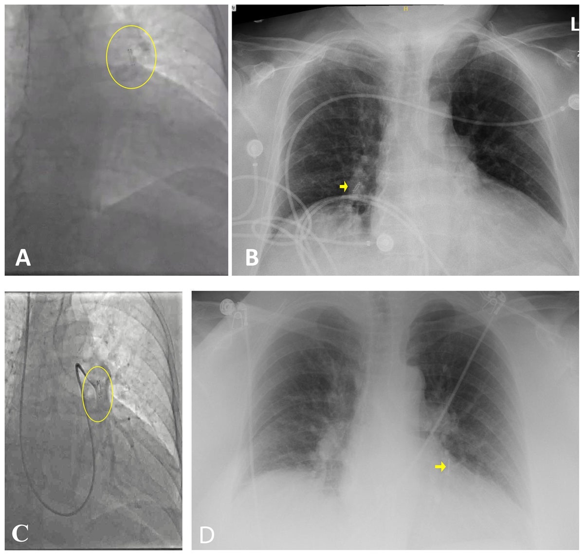
Figure 1
(A) Deployment of the CardioMEMS™ device (Abbott) in a distal left pulmonary artery branch. (B) Post-procedural chest x-ray shows the CardioMEMS™ device in the right middle pulmonary lobe. (C) CardioMEM™ device found re-embolized to the left lobe, in a proximal left pulmonary artery. (D) Chest x-ray shows the CardioMEMS™ device in place in the lower left lobe months after the procedure.
The Swan-Ganz catheter was advanced into the left PA and a platinum plus wire was advanced into the A10 branch. The balloon was inflated in the left PA and the catheter was railed forward to lodge the device more distally (Video 1). Its final position was confirmed to be stable.
Video 1
(A) Repositioning of the CardioMEMS™ device (Abbott) in the left lower pulmonary lobe using a (B) Swan-Ganz catheter pushing the device distally for optimal stable repositioning until (C) final device deployment. Also view Video 1A at https://vimeo.com/1177056731; Video 1B at https://vimeo.com/1177057837; Video 1C at https://vimeo.com/1177058742.
A contingency plan was in place. If repositioning had resulted in PA injury, device instability, or failure to achieve an adequate position, immediate conversion to snare retrieval was planned, with surgical backup available if needed. Continuous fluoroscopic guidance and careful catheter manipulation were used to minimize the risk of vascular trauma throughout the procedure.
Outcome and Follow-up
Three days after the procedure, the patient was discharged in a stable condition with improved dyspnea (NYHA II) and regained mobility. CardioMEMS™ device pressure readings were monitored and reviewed weekly, leading to adjustments in her guideline-directed medical therapy (GDMT), as needed. A chest x-ray was repeated 10 months later and showed the device in a stable position (Figure 1 D).
Discussion
This case represents the occurrence of early embolization of a CardioMEMS™ PA hemodynamic monitoring device despite a successful deployment followed by a second embolization to the same original location. To ensure future stability, the device was pushed forward using a balloon-tipped catheter.
Heart failure is an escalating global public health concern with increasing incidence worldwide.2 The CardioMEMS™ HF System compromises a wireless sensor implanted in a branch of the left PA. This sensor employs microelectromechanical system (MEMS) technology and a piezoelectrical membrane to measure PA pressures.3 In certain HF cases, the CardioMEMS™ device is advantageous for managing both high and low filling pressures, reducing hospitalization rates in patients with NYHA Class III, and enabling optimal GDMT, with continuous monitoring even after discharge.1,4
The CardioMEMS™ device does not have a traditional anchoring mechanism, which can potentially increase the risk of embolization. This concern has been documented in case reports, but the frequency and commonality of this issue is still low.1,5 Notably, large-scale clinical trials and registries evaluating the CardioMEMS™ system have not systematically reported post-implantation device embolization as a distinct safety end point, with device-related adverse events being captured within composite measures such as device/system-related complications and unanticipated serious accidents. The consistently low rates of these composite safety outcomes across studies further support the rarity of clinically significant embolization events.
Although uncommon, early sensor migration can occur if the device is deployed in an artery larger than the recommended size. Additionally, a suboptimal artery angle can facilitate device migration, especially if deployed in a proximal PA branch. Blood flow direction may also contribute to contralateral displacement of the device. Despite following the implant protocol approved by the Food and Drug Administration—which requires the target site to be in the lower lobe region of either the left or right PA with a vessel diameter of at least 7 mm and an angulation of less than 30 degrees, and positioning the distal loop in a vessel with a diameter between 5 and 8 mm—device embolization can still occur. Notably, this case is distinguished by the device’s migration back to its original implantation site. To mitigate the risk of embolization, it is crucial to adhere to correct procedural techniques and carefully select the appropriate PA branch for deployment.
In this patient, the incidental finding of the CardioMEMS™ migration was concerning because the patient was asymptomatic. This highlights the importance of routine post-procedural chest x-ray monitoring. Addressing this issue proactively could prevent collecting wrong measurements, which could have adversely affected GDMT. Furthermore, in cases of migration, device retrieval is not the only management option; repositioning the device distally with a balloon-tipped catheter may also be effective.
Conclusion
We report a rare complication where the CardioMEMS™ monitoring device can migrate acutely post-implantation despite correct positioning according to protocol followed by self-repositioning into its original location. Repositioning the device using a balloon-tipped catheter to wedge it in a distal location can help stabilize its position.
Summary
This case highlights a rare complication involving the CardioMEMS™ device where, despite initial successful implantation, the device experienced acute migration to the right lower lobe and subsequently returned to its original location in the left lower lobe. This incident underscores the importance of vigilant post-procedural monitoring since device migration can occur even when implantation protocols are followed. Effective management strategies, such as using a balloon-tipped catheter for repositioning, can stabilize the device in a more distal and secure location. Routine chest x-rays and adherence to precise procedural techniques are crucial to ensure device stability and optimal patient outcomes.
Key Points
CardioMEMS™ device migration can occur acutely despite technically successful implantation, highlighting the need for vigilant post-procedural surveillance, including routine chest radiography.
Endovascular repositioning with a balloon-tipped catheter is a feasible and effective strategy to stabilize a migrated device in a more distal and secure pulmonary artery branch, optimizing long-term device performance and patient outcomes.