
Introduction
Infective endocarditis (IE) remains a significant cause of morbidity and mortality, with an annual incidence of approximately 3 to 10 cases per 100,000 person-years in industrialized nations.1 While IE traditionally involves the cardiac valves, nonvalvular endocarditis can affect various intracardiac structures. The Chiari network is a fenestrated web-like structure in the right atrium, representing a remnant of the right valve of the sinus venosus. Present in approximately 2% to 3% of the general adult population, it is typically considered a benign anatomical variant.2
Reports of Chiari network involvement in infective endocarditis are exceedingly rare in the medical literature. In most documented cases, the Chiari network is involved as an extension of infection from adjacent structures or as a secondary site. Our case is distinctive and educationally valuable as it presents infective endocarditis developing solely within a Chiari network, with no involvement of cardiac valves or other intracardiac structures.
Case Description
We present a case of a 52-year-old male with a past medical history significant for hypertension, bipolar disorder, gout, intravenous drug use, and human immunodeficiency virus (HIV) who presented to the emergency department with complaints of left foot pain and lower back pain. The left foot pain started 3 days prior, while the back pain had been chronic.
On arrival, his maximum temperature was 100.5°F, and initial labs showed a white blood cell count of 11.9 × 10⁹/L (normal range 5-10 × 109/L). Electrocardiogram showed normal sinus rhythm with no evidence of acute ischemia, infarction, or conduction block (Figure 1). Blood cultures drawn at that time grew methicillin-sensitive Staphylococcus aureus (MSSA), prompting an infectious disease consult.
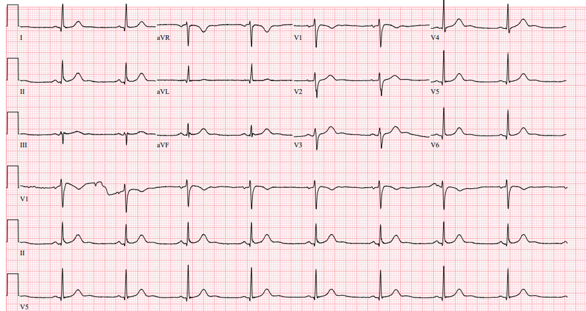
Figure 1
Electrocardiogram showing normal sinus rhythm with no evidence of acute ischemia, infarction, or conduction block.
The patient denied recent injection site infections and had no indwelling prosthetic material. Examination of the right foot revealed swelling, erythema, and pain consistent with a prior gout flare. Magnetic resonance imaging (MRI) of the right foot showed no evidence of osteomyelitis, and symptoms improved with conservative management of gout. His chronic back pain was attributed to degenerative disc changes and gait compensation. Physical examination did not reveal classic signs of endocarditis (petechiae, splenomegaly, heart murmur, Osler’s nodes, or Janeway lesions). No spine tenderness was noted. Given MSSA bacteremia and persistent back pain, echocardiographic and lumbar spine imaging were obtained. MRI of the lumbar spine showed no concern for abscess formation.
A transesophageal echocardiogram (TEE) was obtained to evaluate for intracardiac vegetations. TEE demonstrated normal biventricular function with a left ventricular ejection fraction of 55% to 60% and no significant valvular abnormalities aside from trace regurgitation. No patent foramen ovale (PFO), septal aneurysm, or atrial septal redundancy was seen on TEE. A mobile echo density measuring 1.4 × 0.6 cm was visualized, attached to a Chiari network—an embryologic remnant in the right atrium (Videos 1, 2). These findings led to the diagnosis of Chiari network infective endocarditis.
Video 1
Transesophageal echocardiography in the transgastric right ventricular inflow view revealed an oscillating vegetation in the upper left quadrant of the image; see also at https://youtu.be/zFPVRtzZ1vI.
Video 2
Transesophageal echocardiography in mid-esophageal view showing Chiari endocarditis; see also at https://youtu.be/zv0xCRI2VLU.
The patient was started on intravenous (IV) cefazolin following bacteremia clearance and was discharged with a 6-week course of IV cefazolin after peripherally inserted central catheter placement. The patient was lost to follow-up appointments and did not have repeat echocardiographic imaging to determine resolution.
Discussion
A Chiari network is a mobile, web-like structure of fibers in the right atrium. It originates from the region of the eustachian and thebesian valves at the orifice of the inferior vena cava, with attachments to the upper wall of the right atrium or atrial septum. It is present in 2% to 3% of normal hearts. Usually, it does not hold clinical significance but sometimes can be confused with vegetation, thrombus, tumoral mass, or tricuspid valve disruption.3
Very few cases of a Chiari network associated with infective endocarditis have been reported in the literature. Additionally, no involvement of the valves is a rare phenomenon. There have been a few cases of nonbacterial endocarditis related to the Chiari network. Chiari network has been associated with PFO with more right-to-left shunt than controls, facilitating paradoxical embolism,4,5,6 atrial septal aneurysm, and recurrent cardiac embolic events. Also, it has been associated with supraventricular tachyarrhythmias.7
Our patient is unique in the sense that there were no classic physical signs, dermatologic features, or acute complications related to Chiari network endocarditis as typically described in the literature. Our patient had multiple risk factors for infective endocarditis, especially active IV drug use, and HIV infection. Presentation with nonspecific symptoms of back pain and joint pain rather than classical symptoms of IE, such as fever, murmur, or peripheral stigmata, requires a high index of suspicion for IE in a high-risk population. Transthoracic echo and TEE both are considered reliable tools for diagnosing Chiari network; however, in most cases, TEE was required for confirmation.4,8,9
There are no clear guidelines for the management of Chiari network endocarditis based on size or extent of the vegetation. According to the few existing case reports,4,8 in most cases, complete resolution was achieved with prolonged antibiotic therapy of 6 to 8 weeks supported by follow-up imaging and resolution of symptoms. In some cases, surgical options were considered due to the risk of persistent infection and thrombus formation. In one case report, the patient underwent a surgical approach to a patient who already had another indication for cardiac surgery (three-vessel disease requiring coronary artery bypass grafting).10 Whether these treated patients are at increased risk of another valve infection or thrombotic complications has not been described.
Conclusion
Chiari network endocarditis is a rare and often overlooked cause of infective endocarditis. Its diagnosis can be challenging because it may mimic other intracardiac masses such as thrombus, vegetations, or tumors on imaging. This case highlights the importance of considering Chiari network involvement in patients with unexplained bacteremia and utilizing detailed echocardiographic assessment, especially transesophageal echocardiography, to aid in diagnosis. Early recognition is essential to guide appropriate treatment and to improve clinical outcomes.
Competing Interests
The authors have no competing interests to declare.