
Case Presentation
A 36-year-old woman presented for evaluation of exertional chest pain intermittently over a month and experienced an acute episode lasting 3 hours, accompanied by diaphoresis and dyspnea. She has a medical history of treated Hodgkin’s lymphoma, for which 10 years earlier she had received multimodal therapy including 41 Gy of mediastinal radiation therapy (RT) following chemotherapy. She had no traditional cardiovascular risk factors. A 12-lead electrocardiogram (ECG) showed ST-segment elevation in lead aVR, with ST depression in V1 through V6—a pattern suggestive of obstructive CAD. Selective coronary angiography showed 90% stenosis of the distal left main coronary artery extending into its bifurcation; the right coronary artery was normal (Figure 1, left). Intravascular ultrasound (IVUS) showed diffuse, eccentric fibrous plaque without lipid core or calcification, inconsistent with typical atherosclerosis (Figure 1, right). The patient underwent percutaneous coronary intervention (PCI) with a drug-eluting stent (DES). The patient was started on appropriate medical therapy and discharged a few days post-procedure. She reported no symptoms and good exercise tolerance at a 3-month follow-up.
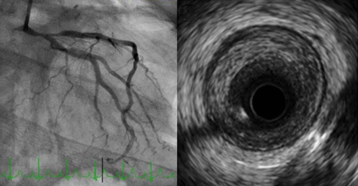
Figure 1
(Left) Coronary angiogram showing severe stenosis of the distal left main coronary artery that extends into the proximal left anterior descending artery. (Right) Intravascular ultrasound image of the distal left main coronary artery showing significant stenosis with circumferential fibrotic plaque without calcification.
Introduction
Radiation-induced coronary artery disease (RICAD) is a serious late complication of chest RT, particularly in survivors of Hodgkin’s lymphoma and breast cancer. It often emerges years to decades after exposure—typically 10 years in breast cancer patients and 2 to 3 decades in Hodgkin’s survivors. The risk is significantly elevated, with increases up to 250% in Hodgkin’s survivors and 25% in breast cancer survivors compared to unexposed populations. Key risk factors include younger age at exposure (< 25 years), higher cumulative RT doses (> 25 Gy mean heart dose), larger irradiated cardiac volumes, inadequate shielding, preexisting coronary atherosclerosis, the presence of typical cardiovascular and metabolic risk factors, and combination with cardiotoxic chemotherapies (such as androgen deprivation therapy, immune checkpoint inhibitors, and vascular endothelial growth factor inhibitors).1, 2, 3, 4
The pathophysiology of RICAD is distinct from typical atherosclerosis. RT injury leads to endothelial cell loss, chronic inflammation, oxidative stress, and fibrointimal proliferation. Histologically, these plaques are rich in collagen and fibrin, with minimal lipid content.5 Lesions frequently involve the proximal coronary segments, described as long, smooth, concentric and tubular.6 Diagnosis is challenging, as many patients remain asymptomatic or present with nonspecific symptoms. When suspected, noninvasive imaging such as coronary computed tomography angiography (CTA) or invasive coronary angiography is often required to define coronary anatomy. Stress perfusion imaging, especially positron emission tomography, is sensitive for detecting clinical, subclinical, and microvascular disease. Intravascular imaging can further delineate the fibrotic, noncalcified nature of these plaques.7
Management of RICAD is complex due to the increased procedural risks posed by radiation-related tissue damage. Surgical revascularization is often limited by poor healing, calcified conduits, and prior chest surgeries, and associated with increased risks of graft failure, perioperative complications, and increased mortality. As a result, PCI with DES and optimizing medical therapy are the preferred treatment modalities over bypass surgery for left main or triple vessel disease, if technically feasible. Surveillance strategies emphasize early identification and aggressive risk factor modification. For high-risk individuals, evidence supports the use of stress perfusion imaging or coronary CT angiography starting 5 years after RT. Given the insidious nature of RICAD, clinicians should maintain a high index of suspicion and adopt a proactive, individualized approach to monitoring and intervention. Finally, a thorough evaluation for valvular, myocardial, pericardial, and conduction pathology due to RT should be performed on all patients prior to intervention, as personalizing treatment decisions utilizing the heart team approach is vital in this complex patient population.7,8
Points to Remember
Radiation-induced Coronary Artery Disease: Risk and Prevention
Patients who have undergone chest RT radiation therapy, especially for conditions like lymphoma or breast cancer, are at increased risk for developing CAD or accelerating preexisting CAD years and decades after treatment. The risk is dose dependent (highest risk for > 25 Gy mean heart dose) and highest when exposure occurs at a younger age (< 25 years). Although RICAD can occur independently of traditional risk factors, aggressive risk factor control offers the greatest potential for reducing the incidence and progression of RICAD. Therefore, preventive care should begin before RT initiation and continue indefinitely.9,10
RT delivery techniques to reduce cardiac radiation exposure should lead to a considerable reduction in risk, including heart-sparing RT strategies, lower intensity RT technologies, the use of deep inspiration or respiratory-gated techniques, and image-guided RT.7,8
Pathophysiology of Radiation-Induced CAD
Radiation can cause endothelial damage, leading to accelerated atherosclerosis, fibrosis, and obstructive CAD. This process often affects the ostial and proximal segments of coronary arteries and can lead to triple-vessel CAD. Intravascular imaging usually shows fibrotic, noncalcified plaque with negative remodeling.
Long-term Surveillance
Given the delayed onset of radiation-induced CAD, long-term cardiovascular monitoring is essential. Survivors of thoracic radiation should undergo periodic cardiovascular assessments, even in the absence of symptoms.
Screening for RICAD should be considered in high-risk patients who have received chest RT in the form of functional imaging and/or coronary CTA beginning 5 years after RT. Functional cardiac imaging should be considered in asymptomatic patients with preexisting CAD or when new significant CAD is detected on anatomical imaging. In asymptomatic patients with inducible ischemia secondary to RT-induced CAD, a heart-team approach is recommended to discuss revascularization options.11
Diagnosis of RICAD
Diagnosing RICAD is challenging due to its delayed onset and variable presentation, and many patients present atypically. Chest pain and dyspnea can also stem from radiation injury to the pericardium, lungs, or chest wall, making diagnosis complex. Therefore, clinicians must maintain a high index of suspicion in patients with prior chest RT.
Ischemic evaluation in patients with suspected RICAD is similar to the general population. Non-invasive imaging modalities like coronary CTA and stress perfusion imaging testing are valuable tools for detecting clinical CAD. Invasive coronary angiography may be warranted based on non-invasive findings and clinical presentation. positron emission tomography myocardial perfusion imaging can be considered for microvascular disease evaluation.
Management Strategies
A multidisciplinary team discussion is highly recommended for clinical decision-making in patients with RICAD and inducible ischemia, left main CAD, or other concomitant RT-induced cardiovascular involvement.11
Treatment of RICAD aligns with standard CAD management, including lifestyle modification, pharmacotherapy, and PCI with DES, when indicated and technically feasible. RT can complicate surgical bypass intervention due to tissue fibrosis and poor tissue healing and is a less favorable treatment option.
Cancer survivors who received chest RT are at increased risk of acute coronary syndrome and carry worse prognosis; however, clinical presentation can be atypical. Immediate coronary angiography and PCI are associated with improved outcomes.12
Conclusion
RICAD is a significant late complication in patients treated with chest RT. Awareness of this risk, coupled with vigilant long-term surveillance and appropriate diagnostic strategies, is crucial for early detection and management, ultimately improving patient outcomes.
Competing Interests
The author has no competing interests to declare.