
Introduction
While manual catheterization remains the cornerstone of contemporary cardiovascular therapy, it is limited by operator dexterity,1 fatigue,2 prolonged fluoroscopy exposure,3 and procedural complications including vessel perforation,4 arterial dissection,5 and suboptimal catheter control.6 Occupational hazards range from musculoskeletal strain7 and radiation-induced skin conditions8 to cataracts,9 carcinogenic effects, and increased rates of solid brain tumors.10 For the patient, other than the ionizing radiation, contrast-induced nephropathy is also a concern during extended procedures.11
In an effort to address these limitations, robotic catheterization systems have emerged as transformative technologies12 aiming to enhance procedural precision, safety, and consistency, with examples such as the CorPath® GRX (Corindus, Siemens Healthineers), Magellan™ (Hansen Medical, Auris Health/Johnson & Johnson), and Niobe® Remote Magnetic Navigation (RMN) (Stereotaxis), each leveraging unique technologies. These platforms have been utilized for various cardiovascular and neurovascular interventions with the goal of addressing challenges regarding control and navigation of conventional manual devices as well as ergonomic and safety issues such as continuous exposure of staff and clinicians to ionizing radiation from X-ray fluoroscopy typically used to guide such procedures.
Robotic systems have been applied in a range of therapeutic interventions, including (1) percutaneous coronary intervention (PCI), with the goal of mechanically restoring blood flow in stenotic or occluded coronary arteries typically via balloon angioplasty (over a guidewire) and often stent deployment; (2) peripheral artery angioplasty and stenting, which are part of the broader category of peripheral vascular interventions (PVI), with the goal of restoring blood flow in the legs and other peripheral arteries; (3) electrophysiology applications and specifically cardiac catheter ablation, with the goal of isolating the arrhythmogenic source from the rest of the myocardial tissue to restore normal cardiac rhythm; (4) transcatheter mitral valve repair or replacement (TMVR/TEER), which involves percutaneous transseptal delivery of devices to repair/replace the mitral valve; (5) transcatheter aortic valve implantation/repair (TAVI/TAVR) for valve replacement or repair for aortic stenosis; (6) and endovascular aneurysm repair (EVAR/TEVAR) for endograft placement to manage aortic aneurysms. Specifically, in neurovascular applications, the primary applications explored have been mechanical thrombectomy, for the removal of thrombus to restore blood flow in acute ischemic stroke patients, as well as aneurysm treatment. All of these interventions span diverse anatomical regions and mechanical demands, motivating the development of robotic systems tailored to specific clinical contexts.
This review systematically analyses 19 robotic catheter systems, categorizing them based on their actuation mechanisms (Table 1; Figures 1, 2), and examines their medical applications and specifications, such as degrees of freedom (DOF), feedback and navigation mechanisms, device compatibility and commercialization phase, and strengths and limitations.
Table 1
Summary of robotic catheter systems, grouped by actuation mechanism, showing degrees of freedom, catheter driving method, tip articulation, feedback modality, compatible devices, procedural focus, and development status. DOF: degrees of freedom; PCI: percutaneous coronary intervention; PVI: peripheral vascular intervention; PNI: peripheral neurovascular intervention; TMVR: transcatheter mitral valve repair
| ROBOTIC SYSTEM | DOF | DRIVE MECHANISM | TIP ARTICULATION | FEEDBACK | COMPATIBLE DEVICES | APPLICATIONS |
|---|---|---|---|---|---|---|
| Mechanical Systems: Roller-Engaged Platforms | ||||||
| CorPath Systems (Commercial) | 2 & 5 | Friction rollers | None | Visual (X-ray with 3D visualization) | 5F-7F catheter, 0.014” guidewire | PCI, PVI, PNI |
| Sentante (Pre-commercial) | – | Friction rollers | None | Visual (X-ray), Haptic | 2F–8F catheters, 0.014”–0.038” guidewires | PCI |
| Bi-motional Roller Cartridge robot (Choi et al.) (Pre-commercial) | 3 | Friction rollers | None | Visual (X-ray), haptic | Standard catheters and guidewires | PCI |
| Robotic System for Endovascular Surgery (ROSES) (Pre-commercial) | 17 | Friction rollers | None | Visual (X-ray) | 2F-20F catheters and 0.014” guidewires | PCI |
| Liberty (Pre-commercial) | – | Likely Friction rollers | None | Visual (X-ray) | 4F-6F catheters, 2F-3F micro-catheters, 0.014”–0.018” guidewires | PVI |
| Vascular Interventional Robot (VIR-2) (Pre-commercial) | 2 | Friction rollers | None | Visual (X-ray with 3D visualization), haptic | 4F-6F catheters and standard guidewires | PVI, PNI |
| Mechanical Systems: Gripper-Driven Platforms | ||||||
| Robotic Endovascular Intervention system (REIS) (Pre-commercial) | 12 | Four grippers on guide rail with wire-rope assembly | None | Visual (X-ray) | Standard catheters and guidewires, All cylindrical tools between 0.43 mm-40 mm | PVI |
| R-One (Commercial) | 2 | Mechanical pads | None | Visual (X-ray) | Standard catheters and guidewires | PCI |
| Remote-Controlled Vascular Interventional Robot (RVIR) (Pre-commercial) | 2 | Two grippers on linear guide | None | Visual (X-ray), haptic | Standard catheters and guidewires | PNI |
| Mechanical Systems: Tendon-Driven Platforms | ||||||
| Magellan (Commercial) | 2 | Sliding rollers | Pull-wires | Visual (X-ray) | Proprietary 9F and 6F co-axial sheath and guide catheter, 0.014”–0.035” guidewires | PVI, PNI |
| Sensei/Sensei X (Commercial) | 2 | Linear guide | Pull-wires | Visual (X-ray) | Proprietary 10.5F and 14F co-axial sheath and guide-catheters, < 8.5F ablation catheter | Cardiac ablation |
| Amigo (Commercial) | 2 | Friction rollers | Pull-wires | Visual (X-ray with 3D visualization) | Standard ablation and mapping catheters | Cardiac ablation |
| Capstan (Pre-commercial) | 3-4 | Friction rollers | Pull-wires | Visual (Likely TEE and X-ray) | – | Mitral repair |
| SurgiPulse (Pre-commercial) | 5-6 | – | Pull-wires | Visual (TEE, X-ray) | Steerable guiding catheter, standard guidewires, MitraClip™ G4 and other similar sized mitral repair devices | Mitral repair |
| Microfluidic Systems | ||||||
| Coral (Pre-commercial) | 2-3 | – | Fluid filled balloon-array | Visual (likely X-ray) | Guidewires, microcatheters, and proprietary Clipper™ tool | Mitral repair |
| Stimuli-Responsive Systems | ||||||
| X-Cath (Pre-commercial) | 2 | Friction rollers | Electroactive polymer | Visual (likely X-ray) | Standard microcatheters and guidewires and may include other additional thrombectomy tools | PVI |
| Magnetic Systems | ||||||
| Stereotaxis Robotic Magnetic Navigation (RMN) System (Commercial) | 3 | V-Drive™ (Linear guide) | Magnetic tip and two external static magnetic fields | Visual (X-ray with 3D visualization) | Magnetically enabled standard ablation, mapping, and diagnostic catheters and new MAGiC™ proprietary catheter | Cardiac ablation, PCI |
| Catheters Guidance Control and Imaging (CGCI) (Commercial) | 3 | Linear guide | Magnetic tip with external eight electromagnets | Visual (X-ray) | Proprietary ablation catheters | Cardiac ablation |
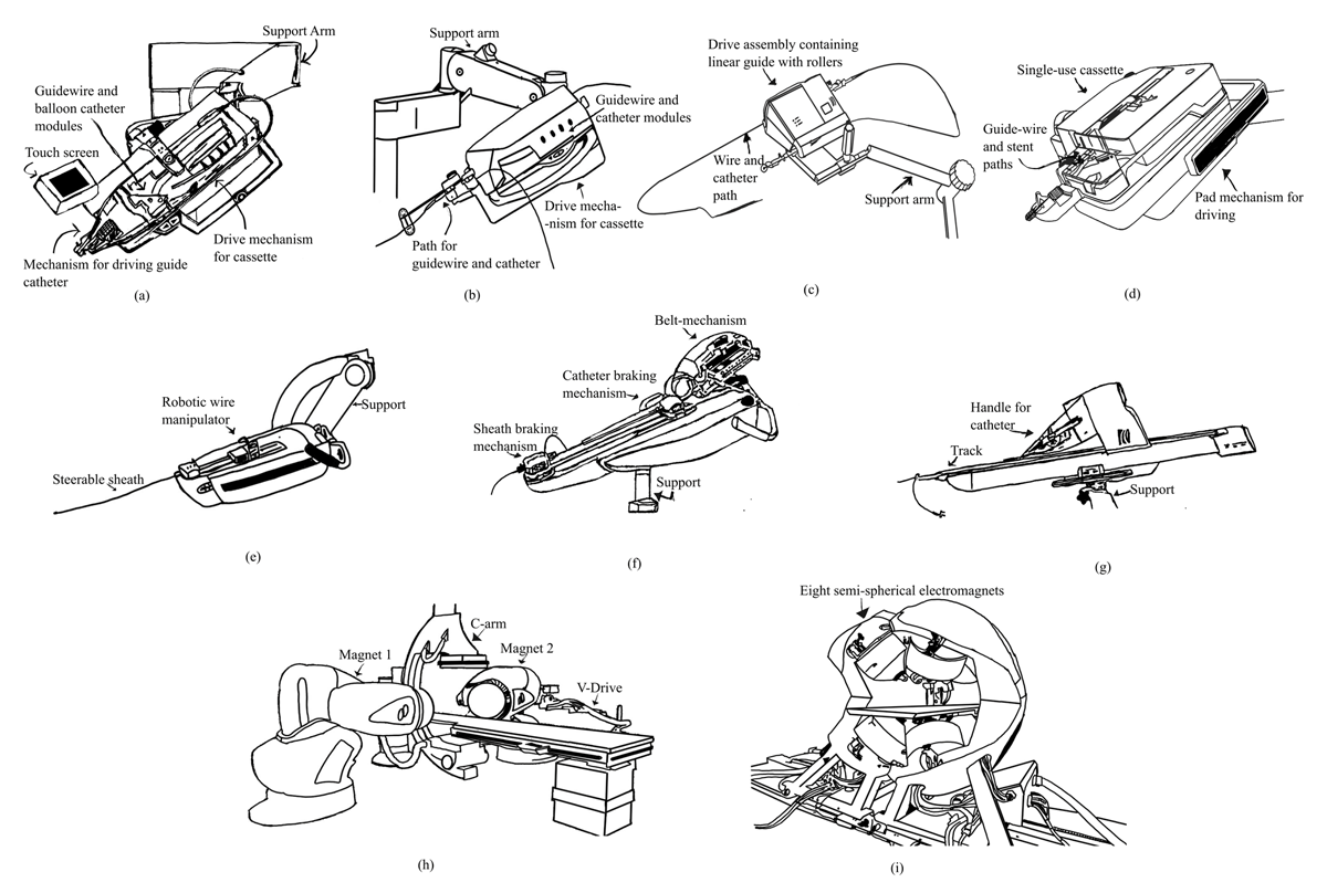
Figure 1
Commercial robotic systems used in endovascular and electrophysiological interventions. (A) CorPath® GRX; (B) CorPath® 200; (C) Liberty®; (D) R-One®; (E) Sensei®; (F) Magellan™; (G) Amigo™; (H) Stereotaxis™ Remote Magnetic Navigation; (I) Catheter Guidance Control and Imaging™
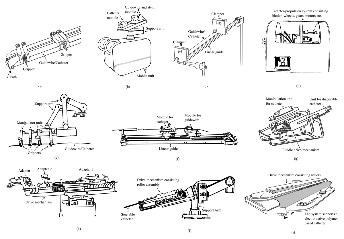
Figure 2
Pre-commercial and investigational robotic systems under academic or industrial development. (A) Sentante™; (B) Bimotional roller cartridge (Choi et al.); (C) Robotic System for Endovascular Surgery; (D) vascular interventional robot; (E) Robotic Endovascular Intervention System; (F) remote-controlled vascular interventional robot; (G) Coral™; (H) SurgiPulse; (I) Capstan™; (J) X-Cath™
Scope and Inclusion Criteria
This review includes 19 robotic catheter systems selected based on their demonstrated relevance to cardiovascular interventions and the availability of clinical validation data. Inclusion was limited to platforms that have undergone at least one reported human study—either in a clinical or investigational setting—published or made available up to April 2025. Systems limited to animal studies, simulation-only evaluations, or low technology-readiness levels without demonstrated procedural feasibility were excluded to maintain translational relevance.
Two exceptions to this criterion—Coral™ (Moray Medical) and X-Cath™ (XCath)—were retained due to their novel actuation strategies and imminent commercial readiness, which directly support the review’s emphasis on actuation-based classification. Additionally, historically important but commercially discontinued systems such as Sensei® (Hansen Medical, Auris Health/Johnson & Johnson) and Magellan were included since they have generated substantial clinical data and serve as key benchmarks for evaluating subsequent platforms. Highly conceptual or early-stage prototypes without demonstrated procedural relevance were also omitted. Soft robotic and continuum-based catheter systems, though increasingly explored in academic settings, were excluded as they remain investigational and lack published in vivo human validation.
Mechanical Systems
Mechanical systems quantitatively dominate the field of medical robotics research. For cardiovascular interventions, they typically rely on direct mechanical manipulation of the catheter. Some designs rely on proximal catheter manipulation using actuators like rollers, clampers, and conveyor belts while others enhance motion control by incorporating additional tip or shaft manipulation (eg, through tendon-driven mechanisms).13
Roller-Engaged Platforms
Friction-roller mechanisms are fairly common as they provide a simple yet relatively reliable mechanism for direct proximal manipulation of the catheter, similar to conventional manual operation. The CorPath® family (Corindus, Siemens Healthineers) incorporates this approach. The CorPath 200 utilizes two rollers engaging a single-use sterile cassette to advance a standard 0.014″ guidewire along with a proprietary 5F to 7F catheter. This system was initially developed for PCI14,15,16 and later extended to PVI.17 Despite the widespread clinical use, analysis of the system’s failure modes in numerous PCI studies revealed a lack of guide catheter support as the main reason for manual conversion.18 Such limitations were addressed through the CorPath GRX upgrade, where all three devices (ie, the guidewire, stent/balloon catheter, and guiding catheter) were controllable with the robot, extending its use to neurovascular interventions.19,20,21,22,23 Alongside remote teleoperability,24 the system also allowed for automation of simple common techniques, such as spinning and side-to-side motion of the wire or the Dotter technique (rapid advancing and retracting of the wire).25 However, after acquisition by Siemens Healthineers, the system was discontinued in 2023 despite clinical interest.26
More recently emerging platforms such as the Sentante™ system (UAB Inovatyvi Medicina/Sentante) extend roller-based designs by supporting a broader range of guidewire and catheter sizes and allow simultaneous manipulation of up to three devices.27,28 The system also incorporates force-feedback for the user to allow for haptic interface and promises increased safety. Preliminary in vivo studies and an ongoing, unpublished first-in-human PVI trial (NCT06537947) suggest procedural success, positioning Sentante as a promising candidate for broader adoption pending regulatory clearance based on upcoming clinical validation.29
In parallel, other designs have been explored to address sterility, device interchangeability, and ease of maintenance. Choi et al. introduced a bi-motional roller with an active clamping mechanism to adapt to various device sizes dynamically.30 The design consists of a disposable unit capable of simultaneously advancing and rotating three devices such as guidewires, balloon/stent catheters, and sensor catheters (eg, intravascular ultrasound). Designed to mount rapidly onto a compact drive unit, it replicates surgeon push-pull and wrist rotation via a 3-DOF haptic controller, providing both torque and force feedback.31 In a two-patient right coronary artery trial, the guidewire was navigated entirely from the console without manual handling, eliminating the need for bedside wiring.30
Building on the same modular philosophy, the Robotic System for Endovascular Surgery (ROSES) (Calabrian High Tech-CHT srl),32 alongside its equivalent version for angioplasty,33 incorporates a friction-roller assembly with dual actuators that independently control guidewires and catheters with the operation of a single device at a time. A three-motor gear train drives a disposable unit with synchronized rollers advancing devices ranging from 0.014″ guidewires up to 20F catheters. The design supports quick-release device exchange and motor-step tracking for lesion-length estimation. In a first-in-human PCI, ROSES successfully delivered and deployed a stent using robotic control, with outcomes comparable to conventional procedures.34 Broader clinical validation is ongoing, especially in highly tortuous and calcified anatomies.
Expanding toward full-console architecture, platforms such as the vascular interventional robot (VIR-2) aim for versatility across broad vascular beds.35 VIR-2 uses friction rollers to vary the catheter shaft pressure, allowing flexible advancement while minimizing jitter and backlash. Mounted on a multi-DOF arm, it is operated via a master console replicating manual manipulation, supported by dual-angle imaging for trajectory planning. The system demonstrated feasibility inhuman cerebral angiographic studies36 and has also enabled remote teleoperation.37
Shifting away from larger-footprint console-based robotic architectures, the Liberty™ system (Microbot Medical Inc.) introduces a fully disposable table-side drive unit optimized for 4F to 6F catheters and microcatheters with a mobile hand-held interface.38 Though not explicitly labeled as a roller-based platform, the mechanism functionally aligns with roller-engaged systems using frictional contact to actuate standard instruments without relying on pull wires or proprietary components.39 Early clinical evaluations from the ACCESS-PVI trial have favorably highlighted its rapid deployment and minimized learning curve.40 Liberty stands out for its miniature design and streamlined workflow, reducing capital investments and setup requirements.
Gripper-Driven Platforms
Unlike roller-based platforms that rely on frictional contact while the rollers are rotated—a challenging task, particularly for smaller diameter devices—gripper-driven platforms use clamps, pads, or jaws to physically grasp and lock the device and manipulate them proximally along a guide rail or actuation track. These designs typically mimic the concept of the clinician’s thumb and index finger grasp/release of the device and enable high precision in clamping, torque delivery, and axial control, often leveraging multi-DOF manipulator assemblies and other mechanisms for large sheath access navigation.
The Robotic Endovascular Intervention System (REIS) exemplifies this approach, employing four grippers driven by a wire-rope assembly to deliver 12 DOF that includes the clamping for rotation and linear motion of up to four devices.41 Its comb-shaped end effector accommodates devices from 0.43 mm to 40 mm in diameter, supporting rapid-exchange and over-the-wire devices. In its five-patient first-in-human study for endovascular aortic repair (EVAR), REIS performed guidewire insertion, stent graft deployment, and contralateral gate cannulation.42
Similarly, using proximal manipulation of the devices using pads, R-One™ system (Robocath) introduces a single-cassette, dual-instrument control mechanism that enables independent, bidirectional manipulation of both the guidewire and balloon/stent catheter for PCI applications.43 It is compatible with all commercially available 0.014” guidewires and rapid exchange stent/balloon catheters. To enhance operational safety, the system automatically parks the rollers and engages mechanical brakes when idle or upon fault detection, which also allows for stabilizing the guidewire. Early first-in-human feasibility studies demonstrated promising clinical success,44 although manual conversions remained common due to insufficient guide-catheter support—a limitation noted in case studies of the 2023 R-One+ successor system.43
The Remote-Controlled Vascular Interventional Robot (RVIR) by Bao et al. similarly employs a clamper-driven actuation using a static conical clamp to secure the catheter and a ball-spline mechanism to decouple torque from the axial motion.45 This is facilitated by a guidewire and catheter module. The interventionalist’s input is captured via a haptic interface (with torque and force feedback). The system’s interface accommodates real catheter handles, magnetic brakes, and pantograph-style torque feedback, which allows the user to leverage their gained experience and dexterous skills while having force and torque feedback.46 In a porcine study, the setup employed a 5F femoral sheath, 5F angiographic catheters, 0.035-inch guidewires, and microcatheter–micro-guidewire pairs.47 Early human cerebral angiographic trials also demonstrated effective force transmission and reduced operator exposure to ionizing radiation, as expected.48
Tendon-Driven Platforms
While the forementioned platforms primarily focus on proximal axial and radial manipulation of the catheter shaft, tendon-driven systems extend robotic control by enabling distal tip steering. Pull-wires run along the catheter shaft and anchor at the tip, with actuators proximally adjusting tension to deflect the catheter tip and even control its shape. This approach, combined with the aforementioned axial and radial manipulations, allows for 3-DOF manipulation of the catheter tip. Some designs may accommodate further DOF to allow for the catheter body to conform to desired shapes and paths.
The Hansen Sensei® Robotic Navigation System (Hansen Medical, Auris Health/Johnson & Johnson) exemplified this approach. It was developed for electrophysiological (EP) procedures, particularly atrial fibrillation and ventricular tachycardia ablation.49 It used a 14F outer and 10.5F inner Artisan steerable sheath actuated via pull-wire tendons and was mounted on a patient-side robotic arm. Operators remotely manipulate the system through a 3-dimensional (3D) joystick console integrated with a customized CoHesion software suite, which overlays electroanatomic mapping data from CARTO 3 and NavX/Ensite to enable intuitive 3D navigation. Real-time contact force feedback was provided through an IntelliSense® module that issued visual and vibratory alerts and was further refined in the Sensei X upgrade alongside enhanced control responsiveness. Various clinical studies have reported improved catheter stability, reduced fluoroscopy, durable lesion delivery,50 up to 90% arrhythmia-free survival in atrial fibrillation51 and a 95% reduction in implantable cardioverter-defibrillators therapies for ventricular tachycardia.52 The system’s dual-sheath articulation and force-aware control later informed the development of the Magellan platform.
The Magellan platform was mounted at the end of the fluoroscopy table and the robotic arm advanced a 9F outer sheath and 6F inner catheter coaxially, both steerable via tendon-based tip articulation and capable of full axial rotation. Magellan supported full robotic control of 0.014”, 0.018”, and 0.035” guidewires. Clinical studies in thoracic EVAR demonstrated reduced vessel wall contact and significantly lower peak forces across arch branches—benefits linked to the system’s stable coaxial navigation and programmable auto-retraction. Notably, transcranial Doppler monitoring during arch navigation showed that robotic operation nearly eliminated micro-embolic signals per catheter maneuver compared with manual withdrawal.53 Additional trials in EVAR54 and embolization55,56 for aneurysm repair highlighted the system’s adaptability across anatomies. However, the eventual market withdrawal in 2019 after its acquisition by Auris Health curtailed its wider clinical adoption.57
Along similar lines to platforms developed for mapping and ablation, the Amigo Remote Catheter System (Catheter Precision, Inc.) offers a streamlined approach by externally actuating standard deflectable catheter handles through a bedside robotic cradle.58 Such catheters also use a tendon for tip deflection of the catheter. The platform docks such commercially available catheter handles (eg, Blazer™, EZ STEER) under a sterile drape. Operator input is issued via a handheld remote, which transmits deflection, rotation, and advancement commands to the robotic cradle for total 3DOF. The cradle physically manipulates the catheter handle’s control surfaces to replicate manual operation. This actuation tensions the catheter’s internal pull wires to produce bidirectional tip deflection and axial motion without altering catheter design. The catheter can be redocked or removed at any point without sterility loss. Integrated with CARTO 3 and ThermoCool® SmartTouch® catheters, Amigo supports zero-calibrated force tracking and real-time force time integral for radiofrequency ablation monitoring. Studies report tighter force delivery, reduced fluoroscopy, and no short-term reconnections in cavotricuspid isthmus ablation.59,60,61 However, it is not compatible with cryoablation or a left-sided access approach.58 It must be noted that radiofrequency (RF) ablation for EP had specific requirements for catheter force, contact, and orientation that informed the design of such systems. Given the recent developments in pulse field ablation for EP applications and the significant differences in mechanical engagement requirements, the design criteria and measurables for such robotic systems may require revision, enabling new opportunities.
Expanding tendon-driven actuation into the treatment of mitral regurgitation, the Capstan™ Medical and SurgiPulse Robotics’ solutions were developed for TMVR and transcatheter edge-to-edge repair (TEER), respectively. The Capstan platform uses a multi-spool tendon mechanism to control both steerable catheter segments and implant deployment.62 A distal extension aids anatomical access, while differentially sized rotors apply synchronized tension at both ends of the prosthetic valve for stable, precise expansion. In early 2025, it was used in two TMVR cases and achieved successful positioning and MR resolution,63 although broader outcome data are pending.64
The SurgiPulse (SurgiPulse Robotics Co.) operates with a similar principle to that of the Amigo system, where a task-specific conventional device, in this case the SQ-Kyrin-M® TMVR system, is directly manipulated in all its degrees of freedom by the robot for teleoperation.65 The system features a robotic arm with three adapters to mechanically couple to the interventional device; these adapters have tendon-based deflection capabilities, enabling precise positioning of the valve implant relative to the mitral leaflets. Friction rollers may supplement this to enable linear guide catheter advancement. The system supports integration with the MitraClip G4 and incorporates echocardiography and angiography for imaging, featuring a clinician console that enables remote fluoroscopy-free operation. In its first-in-human TEER case,66 it reduced severe mitral regurgitation to mild in a high-risk patient within 29 minutes with no complications, demonstrating early procedural feasibility.
Microfluidic Systems
As an alternative to tendon-driven systems, microfluidic systems have been explored, which utilize fluid or gas to directly deform or deflect the device of interest, promising to mitigate the challenges associated with cable routing and friction in tendon-driven mechanisms. The Coral™ System (Moray Medical)67 uses a fluid-driven actuation mechanism in which inflation and deflation of balloon actuators within a multi-lumen catheter shaft produce controlled tip articulation.68 The system supports operator-controlled gross movements through the Trident™ interface, with tip articulation executed via hydraulic actuators. The reusable Driver Assembly and Clipper™ toolset includes a steerable delivery catheter preloaded with an edge-to-edge clip. Moray’s Submersion™ software provides procedural control with augmented visualization and with handheld instruments.
Stimuli-Responsive Systems
Stimuli-responsive mechanisms are being increasingly explored for tip actuation in robotic catheter systems, with electropolymeric materials showing promise for fast and efficient articulation. These materials (conductive polymers) undergo reversible volume changes in response to electrical stimuli, enabling active tip deflection, while the remainder of the catheter actuation typically relies on electromechanical components.
Building on this principle, the X-Cath™ system (XCath) features a dual-drive design that independently controls the catheter sheath and a steerable guidewire, enabling precise navigation in complex anatomies.69 The sheath advances smoothly via motor-driven pinch rollers with gear and belt mechanisms.70 At the same time, the guidewire—made from a Nitinol/stainless steel composite—is controlled separately for independent linear and rotational motion. At the tip, an ionic electroactive polymer actuator actively bends the catheter by asymmetric swelling of circumferential electrodes, allowing real-time adjustment of curvature and stiffness. Radiopaque markers ensure fluoroscopic visibility for accurate guidance. This separation of sheath and guidewire control, combined with an actively actuated tip, distinguishes X-Cath from traditional systems relying on single drives or passive tips. The platform’s precise tip articulation improves maneuverability in tortuous vessels, reducing the risk of vessel trauma. Recently, it demonstrated reliable low-latency remote mechanical thrombectomy over 4,000 miles,71 highlighting its responsiveness and potential for teleoperated interventions. Regulatory approval is pending.
Magnetic Systems
Magnetic navigation systems introduce a noncontact method for distal tip steering by adjusting externally applied magnetic fields to directly manipulate magnetized catheter tips without requiring mechanical torque or force transfer through the catheter shaft.72,73 The concept was initially introduced by McNeil for neurosurgery applications74 but was later adopted for cardiovascular and specifically RF ablation in EP, where the need for complex device navigation and accurate control are significant. Typically, to manipulate the magnetic field, either static magnets are manipulated with robotic arms, or dynamic electromagnetic coils are electrically manipulated to achieve the desired field.
The Niobe® Remote Magnetic Navigation (RMN) system (Stereotaxis Inc.) uses opposing permanent magnet pods (0.08-0.12 T) on robotic arms to steer catheters with a magnetic catheter tip within the thorax. The system requires a dedicated electromagnetically shielded room to accommodate safe operation of the robotic system. The latest iteration, GenesisX, features a semi-mobile docking base with integrated magnetic shielding, eliminating the need for a dedicated modified room. The VDrive™ system75 automates catheter advancement and torque control through mechanical coupling and is compatible with several catheters (eg, Webster’s Lasso® and NAVISTAR®).76 The system integrates x-ray fluoroscopy together with management and control through the Odyssey® and Synchrony™ consoles, unifying magnetic navigation, imaging, and mapping—including compatibility with CARTO® RMT and MAGiC™ catheters—and supports intracardiac echocardiography to enhance visualization and maintain stable catheter-tissue contact. Clinically, these systems (ie, Niobe/Genesis) have been shown to excel in complex and restricted anatomies such as pediatric,77 congenital,78 and dextrocardia cases,79 providing precise force-constrained catheter navigation for RF ablation of various arrhythmias.80
An alternative for EP application, which now seems to have been discontinued, was the Catheter Guidance Control and Imaging system (Magnetecs Corporation), which employed eight semispherically arranged electromagnets to generate dynamic magnetic fields with 6DOF for precise catheter navigation.81,82 The system was designed to produce magnetic fields up to 0.14 T with gradients of 0.7 T/m, enabling fine torque and push-pull control. The system integrated fluoroscopy, an Ensite NavX electroanatomic mapping system, and intracardiac echocardiography (ICE) for real-time catheter adjustments and obstacle detection. While system safety and feasibility for lesion formation and successful navigation to desired targets were demonstrated in human studies,83 the system was not adopted commercially, possibly due in part to limitations imposed by the hemispherical positioning of its static magnets, which significantly restricted patient access by clinicians and staff during the operation.
Some other notable magnetic navigation platforms include the Flux One (Flux Robotics), which features a controllable electromagnetic coil mounted on a mobile 7-DOF robotic arm for guidewire manipulation84; Aeon Scientific’s Phocus system (Aeon Scientific AG), which used movable electromagnets for 3D catheter orientation85 but was discontinued despite regulatory approval, possibly attributed to the coils limiting access to the patient; and Nanoflex Robotics (Nanoflex Robotics AG),86 which evolved this technology with a far more compact bedside magnetic field generator and control console and primarily targeting neurovascular interventions for its initial indication. Spanning from phantom validation to early clinical use, these systems represent the forefront of magnetic navigation.
Imaging
X-ray fluoroscopy, often in conjunction with potentially nephrotoxic contrast agents, is the clinical standard for guiding catheter-based procedures due to its real-time imaging, high spatial resolution, and economic efficiency.12,87 Robotic systems have often been integrated with fluoroscopy systems (eg, Niobe™/Genesis™, Corpath® GRX, etc) to enable precise device navigation.88 However, 2D projection fluoroscopy has challenges due to ionizing radiation, limited soft tissue contrast, and limited visualization in complex 3D anatomy.89 Telerobotic systems sufficiently address the issue of ionizing radiation, particularly for staff and clinicians, but other limitations persist. Despite these drawbacks, the compatibility of fluoroscopy with real-time robotic control, including automated path planning and force feedback, continues to make it central to endovascular robotics, particularly as teleoperation helps mitigate the ionizing radiation for clinicians and staff.88 The fundamental limitations of fluoroscopy in tissue characterization, lack of 3D visualization, and the inability to quantify or observe therapy delivery pushes the need for alternative imaging modalities.
Magnetic resonance imaging (MRI) offers soft tissue contrast adjustment, 3D visualization, and functional imaging capabilities without the use of ionizing radiation, making it an attractive alternative to fluoroscopy.90,91 However, the incompatibility of magnetic devices and various robotic technologies makes them unsuitable for guiding robotic-assisted interventions. While MRI-compatible telerobotic catheter navigation systems have been developed and validated in preclinical settings,92 MRI imaging currently is primarily used for preprocedural planning, including anatomical mapping, tissue characterization, and functional assessment, all of which may be potentially further leveraged by robotic platforms for semiautomation of tasks, guidance of therapy, and user assistance; however, due to these limitations as well as cost, the adoption of MRI for intraoperative cardiovascular interventions continues to face significant challenges.88 In contrast, ultrasound offers real-time imaging, portability, cost efficiency, and compatibility with a wide range of robotic and interventional platforms, making it a versatile modality in both diagnostic and intraoperative settings.93
The integration of ultrasound imaging modalities—such as ICE,94 transesophageal echocardiography (TEE),95 and IVUS96—with robotic solutions for cardiovascular applications appears to hold significant potential. ICE provides real-time, operator-controlled visualization of intracardiac anatomy and is particularly valuable during robotic electrophysiology procedures, such as atrial fibrillation ablation and transseptal access, enabling accurate catheter placement and lesion monitoring.97 TEE, especially in its 3D form and in conjunction with robotic-assisted structural heart interventions,98 holds significant potential for applications such as mitral valve repair or closure of septal defects, offering high-resolution guidance for device deployment. IVUS, on the other hand, is highly synergistic with robotic coronary and peripheral interventions, allowing precise assessment of vessel size, lesion morphology, and stent expansion beyond what fluoroscopy can reveal.96 As robotic systems evolve, the seamless integration of these intraprocedural imaging tools will be essential to achieving fully image-guided, radiation-minimized interventions.
To achieve this, hybrid imaging strategies that combine fluoroscopy with real-time ultrasound (TEE, ICE), IVUS, or CT-derived 3D roadmaps are becoming increasingly common in robotic workflows.99 These systems enhance spatial orientation and tissue visualization, particularly in complex structural heart procedures in which multimodal imaging compensates for the limitations of any single modality. In recent developments, artificial intelligence (AI)-enhanced pipelines have been introduced to automatically segment vasculature, generate dynamic roadmaps, and fuse multimodal data in real time, enabling context-aware robotic navigation and adaptive guidance. Recent developments in deep learning models have enabled simultaneous 3D force estimation from dual-fluoroscopic imaging, with H-Net being one such example.100
Despite these advances, challenges such as image latency and motion effects, intermodality misregistration, and the computational burden of deep learning inference remain obstacles to seamless integration.99 Nevertheless, these techniques mark an important step toward robotic systems that operate with greater autonomy and anatomical awareness, laying the foundation for precision-targeted, image-driven interventions.
Force Feedback
In conventional manual cardiovascular interventions, long and flexible devices are manipulated proximally from outside the patient’s body, engaging with the long and tortuous vascular anatomy along their length. These mechanical constraints limit control over the device tip and make accurate and reliable navigation and engagement with the target anatomy highly challenging.98,101 However, interventionalists gain experience in interpreting and utilizing the subtle forces and torque they sense manually to better guide the devices and engage with target tissue. At the same time, in robotic systems in general, even mature commercialized systems lack integrated force feedback, thereby continuing to risk perforation or tissue damage.13,102 These incidents can precipitate acute thromboembolism, vessel restenosis, cardiac perforation, transesophageal injury, or pericardial tamponade, consequently extending procedure times and diminishing the potential advantages robotic assistance offers.13,103 Furthermore, inefficient target tissue contact and engagement can reduce the efficacy of therapy and risk clinical outcomes (eg, in RF ablation).
For robotic systems, operator interfaces predominantly remain limited to devices such as computer mice, joysticks, or buttons devoid of genuine haptic feedback.13,88 Off-the-shelf multi-DOF haptic interfaces (eg, Novint Falcon, Force Dimension Omega series, and Geomagic Touch/Touch X stylus manipulators) have been explored extensively as haptic interfaces for various robotic systems such as VIR104 and RVIR.48 However, such implementations alter the conventional interaction between the clinician and interventional devices and do not capture their dexterous skills in manipulating conventional devices.87 Clinicians are trained with conventional devices and interpreting the subtle forces/torques they correspondingly sense. Robotic solutions have aimed to accommodate this requirement and allow the user to interact with models of catheters/guidewires in the user interface system while reflecting the forces and torques experienced by the main robot.105
Several strategies have been developed to enable force feedback in robotic systems and are broadly categorized into sensor-based, model-based, and image-based approaches.87 Sensor-based force estimation directly measures contact forces using embedded devices such as fiber Bragg gratings or quartz-based sensors.106 One widely used system, TactiCath™ (Abbott), integrates a fiberoptic sensor to monitor axial and lateral contact forces and is fully integrated into robotic workflows like Genesis™ RMN. Similarly, SmartTouch™ employs a spring-loaded tip with embedded coils to visualize contact force and ablation parameters via the CARTO system—offering a graphical interface that facilitates consistent lesion formation.
Model-based approaches estimate tip–tissue interaction forces by interpreting catheter deflection, curvature, or impedance through kinematic or dynamic models.107,108 A notable example is IntelliSense, a multi-body modeling framework that infers contact force from tip curvature. When integrated with the Sensei X™ system, IntelliSense has been shown to guide effective ablation by generating transmural lesions using combined visual and vibratory cues.
Image-based force estimation is an emerging category that leverages deep learning to infer interaction forces from catheter motion and tissue deformation in real-time. For example, H-Net100 employs a multitask neural architecture to perform simultaneous 3D force estimation and stereo semantic segmentation directly from endoscopic video. By removing the need for embedded sensors, this approach supports the development of sensorless robotic catheters and has shown strong accuracy in ex vivo beating heart experiments using fiber Bragg gratings-based ground truth.
In brief, sensor-based methods provide direct measurement but require tight hardware integration, model-based approaches rely on accurate assumptions about catheter mechanics, and image-based techniques, while promising for sensorless robotics, remain computationally intensive and largely preclinical. Hybrid strategies that combine these strengths may offer a more robust path toward reliable real-time force feedback in robotic navigation.
Beyond this technical classification, the clinical replication of these methods is crucial, not only to validate their utility but also to leverage the experience gained from device-tissue interactions. Accurately interpreting the resulting device kinematics and dynamics is essential for understanding how robotic systems engage with target structure. However, given that cardiovascular devices are highly flexible and engage with the tortuous anatomy with significant friction, the actual forces that the devices experience are highly subtle, and considerable experience is required by the clinician to interpret them.105 Therefore, achieving true closed-loop haptics necessitates substantial enhancements in both sensing accuracy and actuation responsiveness on the patient and operator ends.87 On the algorithm front, the closed-loop force systems typically lack a guarantee of global stability and are prone to system delay issues or operator/device engagement/disengagement. While work has been carried out to utilize environment impedance estimation, instead of directly measured force reflection, such efforts are yet to be transferred to the clinical settings for robotic cardiovascular navigation systems (RCNS) systems.109,102,110
Economics
The economics of endovascular robotic procedures encompass substantial fixed and variable costs shaped by capital investments, maintenance fees, and preprocedural expenditures. Among widely adopted systems, CorPath GRX, primarily used for PCI, demands an initial capital investment that was estimated to be approximately $500,000 to $600,000 USD.111,112 Each procedure incurs additional costs from single-use cassettes (~$700), guidewires, balloons, stents, and catheters, the selection of which depends on lesion complexity. Moreover, pre-procedural angiograms for lesion assessment further raise overall costs. In the context of carotid artery stenting, reports indicate higher overall costs with CorPath GRX due to elevated personnel and supply demands.22 Despite initial uptake, the system was discontinued in 2023,26 underscoring the difficulty of sustaining capital-intensive models, potentially due to limited clinical scalability and workflow compatibility.
The Magellan™ robotic system carried a capital cost estimated at approximately $600K USD, comparable to GRX, but potentially entailed higher cost for the disposables.112 Although clinically validated in EVAR and embolization, Magellan was also withdrawn from the market after its acquisition by Auris Health in 2016,57 potentially reflecting similar commercial challenges.
In catheter ablation, the Stereotaxis® remote magnetic navigation systems present a different economic model. An upfront purchase and installation cost of approximately $2M USD is typically required, with annual maintenance costs exceeding $100K USD.113 Other costs per procedure include the need for specific dedicated proprietary ablation and mapping catheters, among others, depending on procedure complexity and clinician preference, which may further add to procedural costs.113 Given these costs, RMN-guided ablation can potentially benefit from economic efficiency through its reduced capital costs, expansion to cover more procedures, accommodation and compatibility with more off-the-shelf devices, and enabling of new therapies. Despite these costs, the unique capabilities of RMN systems, in precise direct manipulation of the device tip, continue to be unmatched by alternative technologies.
In contrast, emerging systems such as Liberty™ from Microbot significantly shift the economic model by adopting a fully single-use disposable robotic drive, eliminating the need for any initial capital cost investment.114 Comprehensive cost comparisons across platforms remain scarce, in part due to limited public disclosure. Nonetheless, annual financial reports and investor filings offer valuable insight for extrapolating economic viability. It is important to note that most existing economic evaluations do not account for indirect patient-borne charges such as medical staff fees, anesthesia, hospital admission, and postoperative follow-up.
Discussion
Quantitatively, most platforms continue to rely on mechanical roller- or gripper-based drive mechanisms for catheter and guidewire control due to their compactness, cost efficiency, and operational simplicity. However, the most significant clinical adaptation and success can be seen in remote magnetic navigation systems, despite their costs,113 due to the potential added clinical value and enablement of control beyond typical manual device operation.
For medical robotic systems, merely offering teleoperation does not appear to be sufficient. The key contributing factor to the clinical adoption and commercial success of RCNS systems appears to depend on whether robotic solutions enable clinicians to achieve tasks more effectively or beyond what they can achieve manually with conventional tools—or possibly even enable new therapies previously unachievable.115 Increasingly, this includes the capacity to operate across geographic distances, allowing expert intervention without physical co-location.116 These emerging forms of long-distance remote operability go beyond intrinsic master–slave control and introduce new opportunities for expanding vascular care access in underserved or time-critical contexts, such as acute stroke or myocardial infarction.117 Their success, however, remains contingent on robust low-latency network infrastructure118 and security, procedural safety in the absence of tactile feedback, and regulatory support for distributed care delivery.
In addition to enabling new capabilities, ease of integration into existing hospital workflows remains a key determinant of clinical adoption. Large-scale systems such as the Niobe® RMN often require dedicated operating rooms due to their infrastructure and space demands, posing logistical and economic barriers to implementation. In contrast, newer systems such as the Liberty™ robotic platform and the compact bimodal cartridge developed by Choi et al. have been designed for greater mobility and plug-and-play integration, thus reducing setup burden and improving compatibility with diverse clinical environments. However, this is also influenced by the type of intervention for which the system is designed. Future robotic platforms should continue to prioritize modularity, minimal footprint, and ease of deployment to support broader adoption across healthcare systems.
Ultimately, high-quality clinical evidence, ideally in the form of randomized clinical trials, is required to support any claims regarding procedural outcomes and will support clinical adoption. It is anticipated that the future direction of robotic systems in cardiovascular interventions will be shaped by demands for improved procedural outcomes, efficiency, operator and patient safety, and cost-effectiveness.
Future development is expected to advance across three interdependent fronts: imaging integration, hardware evolution, and automation. On the imaging side, fluoroscopy remains dominant due to its cost-effectiveness and real-time capability; however, its limitations in soft tissue contrast have prompted the exploration of alternatives, such as IVUS and MRI.91,96 While promising, the integration of these imaging modalities in RCNS is confined mainly to research environments due to challenges in tool compatibility and workflow integration.88 The utilization of these imaging modalities, particularly by combining detailed preoperative volumetric MR images, real-time ultrasound, and fluoroscopy through algorithmic registration, offers a potential path toward automated, image-based navigation and therapy regulation.90,96,119,120
Hardware innovation will require a shift from purely proximal control to architectures that enable reliable distal manipulation to allow clinicians to overcome their current limitations with conventional manual methods. Current mechanical solutions often fail to accommodate direct tip position control relative to target anatomy, particularly in tortuous anatomy, compromising pushability and precision. Furthermore, nonlinear effects such as insufficient axial stiffness, friction along tortuous paths, and backlash in proximal actuation mechanisms can impair tip precision and force transmission. These issues are rarely quantified in clinical reports, but experimental studies have noted such mechanistic effects like lack of guide-catheter support in systems like CorPath® 200 and R-One™. As next-generation platforms pursue distal or hybrid actuation, targeted evaluations—both benchtop and in vivo—will be necessary to expose and address such mechanical inconsistencies. Expandable cable-driven systems121,122 such as those explored by Magellan Biomedical (Richmond Hill) and, on the magnetic navigation front, mobile magnetic navigation platforms such as those under development by Nanoflex Robotics86 and Flux Robotics84 may offer potential solutions, provided they can accommodate procedural flow efficiency and modularity and meet the demands for cost efficiency.
AI and machine learning have significant potential for further application in RCNS.123,124 Real-time analysis and use of biomedical signals, imaging, and sensory feedback can potentially accommodate closed-loop device navigation and, most importantly, therapy delivery. Their role in real-time control, force estimation, and navigation continues to grow, as seen in emerging semiautomated functions, such as the automation of simple technical maneuvers achieved by CorPath GRX.125 With increasing procedural datasets, AI-based algorithms could facilitate closed-loop automation and enable advanced control techniques that permit continuous improvement of diagnosis, procedure planning, and therapy delivery.
Robotic catheter systems are now being conceptually classified from Level 0 (manual with robotic aid) to Level 5 (full procedural automation).126 While most existing systems operate at Level 1 or 2, recent advances in AI-driven segmentation, context-aware motion planning, and data-driven force estimation are enabling task-level autonomy—such as trajectory guidance, valve positioning, and lesion targeting. Though barriers like image latency, generalizability, and regulatory validation persist, these developments signal a shift toward supervised autonomy, where robotic systems act not as replacements but as cognitive extensions. Future RCNS platforms are likely to combine hybrid control architectures with real-time feedback and clinical context-awareness, enhancing procedural safety, precision, and accessibility.
Conclusion
This review examined the current state and emerging directions of robotic cardiovascular navigation systems, with a focus on clinically validated technologies. Various actuation mechanisms, their technical specifications, application strengths, and weaknesses were discussed. The next generation of cardiovascular robotic systems must combine distal control, closed-loop automation, AI-enhanced functionality, and cost-aware design. Achieving this will require collaborative innovation in hardware, imaging, and control interfaces, transforming robotic systems from adjuncts to integral components of interventional workflows.
Key Points
Future robotic systems in cardiovascular interventions must evolve beyond mere teleoperation to offer capabilities that fundamentally enhance procedural outcomes and enable therapies currently unachievable manually, thereby justifying their cost and complexity for widespread clinical adoption.
The integration of real-time, 3-dimensional imaging modalities such as ultrasound and magnetic resonance imaging offers significant potential for advancing robotic navigation toward automated, image-guided interventions that mitigate radiation exposure and improve visualization in complex anatomies.
Hardware innovation must prioritize reliable distal catheter manipulation and precise tip control, coupled with effective closed-loop force feedback, to overcome the limitations of current proximal-only systems and prevent complications such as tissue damage.
To achieve broader market penetration, new robotic platforms should adopt cost-effective designs, such as disposable drive units, and demonstrate enhanced workflow efficiency to address the economic barriers that have led to the discontinuation of previous capital-intensive systems.
The strategic application of artificial intelligence and machine learning will be vital for enabling advanced control techniques, real-time decision support, and, ultimately, semi- or fully automated tasks in robotic cardiovascular interventions, moving toward continuous procedural optimization.
Competing Interests
The authors have no competing interests to declare.