
Introduction
Coronary artery aneurysm (CAA) is a focal dilation of coronary segment that exceeds 1.5 times the diameter of the adjacent normal segment but does not exceed a third of the total vessel length.1 The incidence rate based on coronary angiography varies between 0.3% to 5.3%,2 and it can present as an incidental finding or cause clinical symptoms. Atherosclerotic coronary artery disease and congenital malformations are the leading etiologic factors for development of CAA, followed by local vessel wall injury after coronary interventions, vasculitis, infections, and cocaine abuse.3 Uncomplicated “silent” CAAs are amenable to medical management with antiplatelet and/or antithrombotic agents, whereas rapid growing or symptomatic aneurysmal vessels treatment should be discussed within a heart team for percutaneous coronary intervention (PCI) or surgical intervention.
Case Presentation
A 68-year-old man with a known history of diabetes, hypertension, coronary artery disease, and myocardial infarction (MI) reported subjective fever and chills but denied weight loss or night sweats. He did not have a coronary angiogram during an episode of MI. 15 days later, he presented with shortness of breath and frequent episodes of angina and was admitted to an outside hospital. There, an echocardiogram revealed post-infarction left ventricular posterior wall aneurysm, left ventricular ejection fraction of 48%, and moderate circumferential pericardial effusion with evidence of constriction. The patient underwent pericardiocentesis with removal of 450 mL straw-colored fluid. Cardiac catheterization showed proximal right coronary artery chronic total occlusion, 70% stenosis of proximal left anterior descending (LAD) and diagonal 1 (D1) branch, and 70% stenosis of the obtuse margin branch of the circumflex coronary artery (Figure 1).
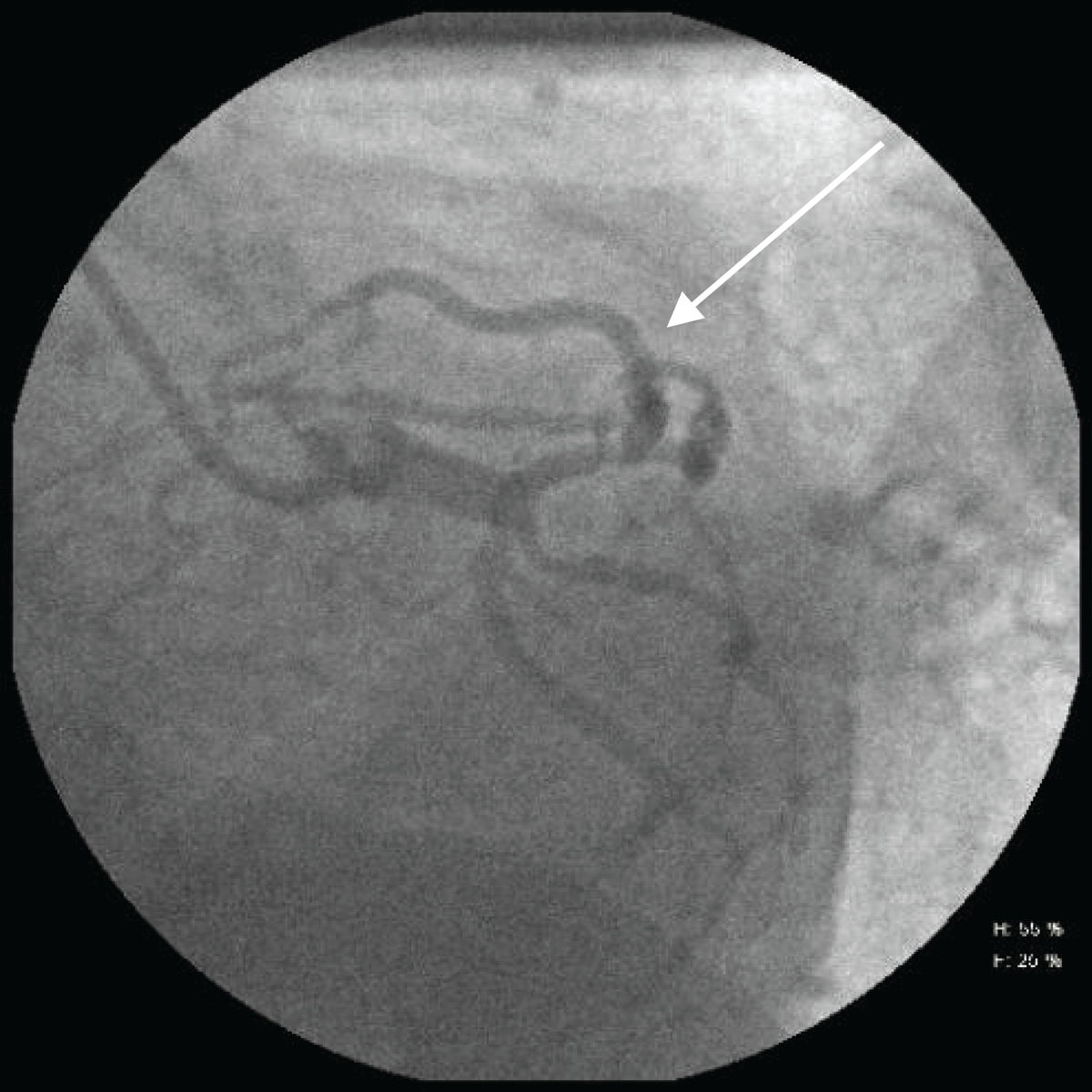
Figure 1
Coronary angiography day 1. White arrow indicates significant stenosis in the proximal left anterior descending artery.
Given the history of multivessel coronary artery disease, the patient was deemed a candidate for coronary artery bypass grafting. After median sternotomy was performed, a visual inspection revealed a thickened pericardium with purulent pericardial fluid and accumulation of fibrin, suggestive of effusive constrictive pericarditis. The cause of pericarditis was unclear based on previous results of a viral infection with a possibility of iatrogenic etiology. Given the thickened pericardium, the bypass graft surgery was canceled and the surgical team proceeded with pericardiectomy and mediastinal washout without revascularization. After surgery and therapy with antibiotics, the pericarditis resolved within 2 weeks, and the patient underwent a new cardiac catheterization 1 month later to address his coronary artery disease. Repeated angiography revealed a coronary artery aneurysm in the proximal LAD and D1 branch bifurcation (Figures 2, 3). The heart team decided to treat the aneurysm percutaneously to avoid redo sternotomy post-pericarditis.
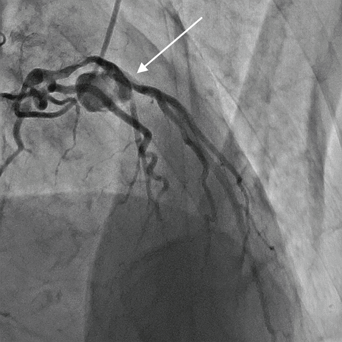
Figure 2
Coronary angiography 1 month after initial presentation. White arrow indicates coronary artery aneurysm in proximal left anterior descending artery and diagonal 1 branch bifurcation.
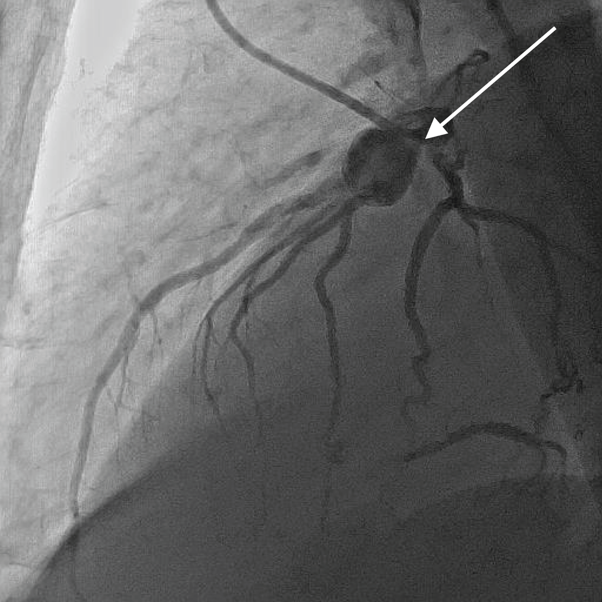
Figure 3
Coronary angiography at 1 month, left anterior oblique 90° view. White arrow indicates coronary artery aneurysm (diameter > 1 cm) in proximal left anterior descending artery and diagonal 1 branch bifurcation.
Percutaneous Coronary Intervention Technique
The LAD artery was initially prepared for stent implantation by predilation using a 3.0 mm × 15 mm noncompliant (NC) balloon (Medtronic NC Euphora™). The balloon was inflated to a pressure of 14 atm to achieve adequate vessel preparation. A covered drug-eluting stent graft was not indicated due to anatomical true bifurcation of the D1 branch of the LAD (1-2.5 mm).
Following predilation, a 3.0 mm × 29 mm MGuard™ stent (Inspire MD) with an integrated polyethylene terephthalate (PET) mesh (initially designed for minimization of distal embolization in acute MI) was deployed in the proximal segment of the LAD. The stent was positioned to ensure full coverage of the diseased segment, including the ostium of the first D1 branch of the LAD.
To optimize stent expansion and apposition within the proximal LAD, the proximal optimization technique was performed using the same 3.0 mm × 15 mm NC balloon, inflated to 16 atm, to expand the proximal portion of the stent against the vessel wall, facilitating improved flow dynamics and side branch access.
Subsequently, our attention was directed to the D1, which was accessed using a Miracle 6™ (Asahi Intecc Co.) stiff wire specifically designed for navigating chronic total occlusions. At the entry point of D1, a balloon predilation was performed using a 2.0 mm × 12 mm balloon inflated to 6 atm to prepare the vessel for adequate opening.
A final “kissing balloon” technique was then used to open both the LAD and D1 branch ostia and carina reposition. This involved simultaneous inflation of a 3.0 mm × 15 mm balloon in the LAD and a 2.0 mm × 12 mm balloon in the D1 branch, both inflated to 8 atm. This step aimed to ensure optimal stent expansion and preserve the patency of the bifurcation.
To complete the procedure, a final proximal optimization was done using the 3.0 mm × 15 mm NC balloon, again inflated to 16 atm in the proximal LAD segment, to ensure ideal stent apposition in the vessel lumen.
Excellent functional and angiographic outcome was achieved, demonstrating complete obliteration of the previously visualized aneurysm with preserved flow in both the LAD and its diagonal branch (Figure 4). The patient was prescribed dual antiplatelet therapy to prevent stent thrombosis along with high-intensity statin therapy to manage atherosclerotic risk and promote plaque stabilization.
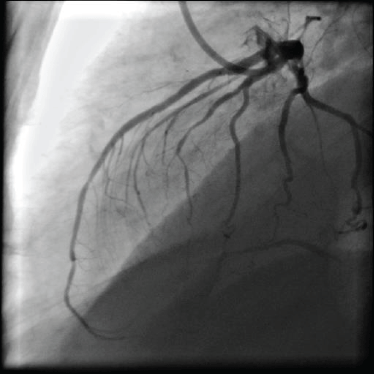
Figure 4
Angiographic result with complete obliteration of the left anterior descending artery coronary artery aneurysm.
Discussion
Coronary artery aneurysms (CAAs) are found in 0.3% to 5% of patients undergoing coronary angiography.1 CAAs can also be detected by magnetic resonance imaging, contrast-enhanced computed tomography, and transesophageal echocardiography.5 CAAs rarely are large enough to be called giant CCA (> 20 mm, or > four-times reference vessel diameter), with the right coronary artery being the most frequently affected.1,2 The earliest medical literature about CAAs (which was issued by the American Medical Association) included observations from case reports that CAAs can “vary in size from that of a pea to that of a pigeon’s egg.”6 Presently, variations of CAAs are more clinically defined and take on saccular or fusiform shapes. Atherosclerotic CAAs are typically fusiform while post-inflammatory CAAs can vary in shape and form. Giant aneurysms are generally classified as saccular and are > 5 cm in diameter.7 The most common site of a CAA is the proximal right coronary artery followed by the proximal LAD, the left main coronary artery,8 the left circumflex artery, and the distal right coronary artery.9 Although inflammation is deemed the fundamental mechanism involved in atherosclerotic aneurysm formation, congenital CAAs comprise nearly 30% of aneurysms in the pediatric population, therefore the exact etiology of CAA development remains unclear.7
CAAs can grow rapidly when associated with pericarditis. While there are a limited number of case reports concerning CAA with pericarditis,2,3 it appears that the interval between the initial symptom and CAA discovery varies from 1 day to 1 year. In most cases, Staphylococcus was found to be causative while systemic inflammatory disease has also been reported.4 In reports involving coronary stent infections, the documented contributory organisms are S. Aureus and Pseudomonas aeruginosa. In theory, a drug-eluting stent may carry a greater risk of infection compared to bare metal stents, considering immunosuppressive properties.9
Conclusions
Coronary artery aneurysm due to a purulent process is a rare complication after pericardiocentesis. Noninvasive cardiac imaging plays a significant role in the early detection of CAAs because underlying complications such as atherosclerosis are usually not suspected. Bacterial identification can also unveil causative agents, therefore antibiotic treatments should be discussed in suspected cases of CAA. Percutaneous coronary intervention is an effective and safe method of treating coronary artery aneurism, especially since infectious cardiac complications after PCI are uncommon. Stents with a polyethylene terephthalate net may be useful compared to a polytetrafluoroethylene graft for CAA in a true anatomical bifurcation, when a large LAD D1 branch is more than 2.5 mm in diameter. A guidewire crossing technique using an MGuard stent is possible when utilizing the stiff chronic total occlusion wire.
Competing Interests
The authors have no competing interests to declare.