
Introduction
Peripheral artery disease (PAD) is a major global health burden, affecting over 200 million individuals worldwide and responsible for significant morbidity, cardiovascular mortality, and healthcare costs. A key factor in PAD management is the accurate visualization and segmentation of vascular plaques and plaque composition. Imaging modalities such as computed tomography angiography and ultrasound (US) are used for plaque assessment in everyday clinical practice. However, they have limited ability to characterize plaque microstructure, particularly in differentiating between soft lipid-rich plaques and hard calcified lesions, for precisely guiding clinical decisions about device selection.
Recent advancements in artificial intelligence (AI) and deep learning-based models have revolutionized many aspects of medical imaging, ranging from image segmentation to automated pathological feature classification. While most of the AI-based vascular plaque analysis tools have been developed with a prime focus on coronary and carotid artery plaques,1,2 there is a marked lack of dedicated tools for lower extremity arterial plaques. Creating such tools is important due to fundamental differences in plaque composition: lower extremity plaques often exhibit greater heterogeneity, higher calcific burden, and more advanced fibrotic remodeling, likely due to differing hemodynamic forces, vessel wall biology, and chronic exposure to peripheral vascular risk factors.
Magnetic resonance imaging (MRI) has emerged as a powerful noninvasive imaging tool for vascular plaque characterization, with excellent soft tissue contrast and multi-parametric imaging capabilities.3,4 Among the MRI techniques, ultrashort echo time (UTE) MRI sequence has the capability of detecting tissues with very short T2 relaxation times to better identify fibrous and calcified areas, which usually cannot be distinguished from each other on conventional MRI sequences.5 Furthermore, UTE-based sequences provide increased detection of signals in heavily calcified regions, which is a major limitation of standard MRI techniques.3 While these advantages are present, UTE MRI remains labor-intensive and susceptible to interobserver variability, necessitating the addition of AI for enhanced diagnostic performance and workflow automatization.
In this review, we first outline current clinical imaging techniques for plaque characterization and examine how AI has been applied to enhance diagnostic performance in PAD. We then present our group’s recent experimental work on a novel MRI-based protocol integrated with AI for lower extremity plaque evaluation, an approach that, even at an early stage, shows clear potential to surpass the diagnostic precision, risk stratification capability, and workflow efficiency of the current standard of care. Ultimately, by bridging advanced imaging physics with AI-driven analytics, this work aims to lay the groundwork for next-generation, precision-guided PAD diagnosis and intervention.
Plaque Formation and Imaging Strategies in Lower Limb Peripheral Artery Disease
Vascular Plaque Pathophysiology
Vascular plaque development is a multifactorial process that begins with endothelial injury and chronic inflammation, resulting in lipid deposition, recruitment of inflammatory cells, and proliferation of smooth muscle cells that cumulatively constitute atheromatous lesions with a high content of lipids, fibrous tissue, and calcific deposits.6 The situation is further complicated in lower limb arteries (particularly in below-the-knee vessel segments) by the frequent occurrence of medial artery calcification (MAC), a process powered mainly by arterial stiffness and hemodynamics changes, most notably seen in diabetic patients or those with impaired renal function.7
MAC is unlike calcified atherosclerotic plaques, which occur in the intimal layer, are a source of luminal narrowing, and directly impair blood flow with associated symptoms such as claudication or critical limb ischemia. Rather, MAC is characterized by calcium deposition in the tunica media without significant luminal compromise but that nonetheless leads to reduced vascular compliance and makes endovascular procedures more difficult.8,9 The recognition of these pathophysiological patterns is essential for accurate diagnosis in order to tailor the most effective revascularization strategies in each individual case, underscoring the critical role of high-resolution vascular plaque imaging in defining plaque morphology and guiding appropriate therapeutic decisions.
Imaging Modalities
Conventional Imaging in PAD
Ultrasound is highly favored for noninvasive imaging due to its real-time hemodynamic evaluation of blood flow, stenosis severity, and plaque morphology. It is particularly useful for identifying soft, fibrotic, and calcified plaques, but heavily calcified vessels and operator dependence can limit its accuracy. It also frequently lacks the resolution necessary to visualize deep or distal segments of the tibial and pedal arteries.10,11 Intravascular ultrasonography is an invasive modality that provides detailed cross-sectional images of the vessel walls, providing data on plaque burden, lumen narrowing, and stent positioning. However, it is costly and requires catheterization, thus it rarely is used outside an interventional setting.12
Digital subtraction angiography (DSA) remains the gold standard for real-time lumenography. However, while it visualizes contrast flow within the vessel lumen, it offers limited or no insight into the arterial wall and does not provide information about plaque morphology, limiting its ability to differentiate the different tissue materials. DSA does provide high-resolution real-time imaging and is often used during endovascular interventions to assess vessel patency and guide vascular access.10,13 On the other hand, it is invasive, requires iodinated contrast agents, and exposes patients to ionizing radiation.
Computed tomography angiography (CTA) is a widely available high-resolution imaging technique with the capability of rapid three-dimensional visualization of the arterial system, making it useful for treatment planning and preoperative evaluation.
Modern multidetector CTA can image from the aortic arch to the feet in one single scan, which significantly speeds up the workflow. However, heavy calcifications can lead to blooming artifacts, deterring its ability to effectively distinguish plaque components. In addition, its diagnostic accuracy in below-the-knee arteries is typically compromised since it is hard to separate dense calcification from intraluminal contrast material in small-caliber vessels.14,15 Photon-counting CT provides better soft tissue contrast and plaque characterization15,16 without the artifacts from heavy calcification and may serve as a promising alternative to standard CTA. However, application remains uncommon in lower-limb extremity arterial disease since no approved protocol currently exists for scanning this region.
Magnetic Resonance Angiography: An MRI-Based Technique for Targeted Visualization of Blood Vessels
MRI has emerged as a powerful noninvasive tool for vascular imaging, with excellent soft tissue contrast without ionizing radiation. In the context where MRI is specifically applied to visualize blood vessels, it is referred to as magnetic resonance angiography (MRA). MRA techniques have been employed in the diagnostic evaluation of PAD, with specific advantages and limitations depending on the clinical context and anatomical region of interest.15 The most common technique used in PAD diagnostics is contrast-enhanced MRA (CE-MRA), which involves intravenous administration of gadolinium-based contrast agents to create high-resolution images of the arterial lumen.17
CE-MRA is widely used for its ability to visualize long arterial segments with high spatial resolution, but it provides limited information in terms of vessel wall characteristics and plaque composition. Additionally, its reliance on gadolinium-based contrast agents limits its use in patients with renal dysfunction. For patients with contraindications to contrast agents, such as those with advanced renal impairment, noncontrast MRA techniques are increasingly utilized.15,17
Quiescent-Inflow Single-Shot MRA is a noncontrast technique that enables rapid, motion-insensitive imaging of the peripheral arteries by capturing inflowing blood during diastole; however, it remains primarily a lumenographic method and lacks plaque characterization capabilities.15,18 Time-of-flight MRA and phase-contrast MRA are other noncontrast methods, although their clinical application in PAD is limited due to lower spatial resolution and longer acquisition times. More advanced vessel wall imaging sequences, including black-blood MRA and multicontrast MRA protocols, are under investigation to evaluate plaque morphology and composition but are not routinely implemented in clinical practice.19,20
The Role of Artificial Intelligence in Vascular Imaging Built on Clinical Imaging Settings
A large amount of recent works demonstrate the growing role of AI-based tools for PAD management in diagnostic, prognostic, and patient-centered domains with the primary focus on early detection, risk prediction, and clinical decision support.21,22,23,24,25,26,27,28 In addition, AI-based tools are becoming essential to enhancing the diagnostic accuracy and efficiency of imaging modalities used in PAD. Among the modalities, the majority of the work is directed towards CTA and US, with comparatively fewer studies on MRA and particularly CE-MRA.
Computed Tomography Angiography
This modality is currently the most frequently used imaging method in AI-supported PAD radiological research. A recent meta-analysis by Jie et al.29 reported that AI-assisted CTA had excellent diagnostic performance in detecting atherosclerotic plaques, with pooled area under the receiver operating characteristic curve (AUROC) values of 0.95 for ≥ 50% stenosis and 0.96 for ≥ 70% stenosis. Similarly, calcifications were detected with high accuracy (AUROC of 0.92). Dai et al.30 also presented important work in this field by proposing a deep learning model, p-EffNet, for above-knee and below-knee stenoses classification (achieved AUCs of 0.987 and 0.981), matching radiologist-level accuracy while significantly increasing efficiency. The p-EffNet uses EfficientNet,31 a type of network that scales convolutional neural networks (CNNs), to classify stenosis degree. Additionally, Mistelbauer et al.32 introduced a semiautomated vessel tracking system that improved segmentation speed by 39%, maintaining excellent accuracy even in the presence of calcifications or imaging artifacts. Their algorithm uses computer vision techniques such as thresholding-based segmentation, filtering, and contour detection to represent vasculature, together with vessel tracking and graph generation. They aim to speed up the creation of large, expert-annotated datasets for machine-learning algorithms that can fully automate these tasks.
Ultrasound
Ultrasound diagnostics have also benefited substantially with AI integration through automated image interpretation, reduction of operator variability, and improved detection accuracy. Luo et al.33 developed a hierarchical deep model that accurately classified lower extremity arterial Doppler waveforms with up to 97% accuracy for normal findings and 88% to 90% for aortoiliac, femoropopliteal, and trifurcation disease levels equaling the diagnostic performance of experienced vascular specialists. They also used other machine learning algorithms such as random forests, support vector machines, and multilayer perceptrons to predict the degree of stenosis. Advances such as the robotized ultrasound system of von Haxthausen et al.34 have demonstrated fully autonomous vessel tracking ability, with a CNN-based image analysis pipeline to autonomously detect and follow vessel lumens. The system achieved vessel visibility in 100% of phantom images and kept the vessel centered with < 4 mm mean absolute error, demonstrating feasibility for autonomous PAD imaging. Moreover, Jiang et al.’s VesNetSCT++ model35 has set new benchmarks in real-time segmentation of femoral and tibial arteries using B-mode and color Doppler US, achieving superior Dice scores over traditional architectures. Their model uses a fully convolutional encoder-decoder backbone to include spatiotemporal context to support B-mode and color Doppler frames.
Magnetic Resonance Angiography
Although relatively limited in number compared to CTA and US, there are also MRA-based studies incorporating AI for the evaluation of PAD—particularly focusing on CE-MRI for assessment of microvascular perfusion and skeletal muscle tissue characteristics of the lower extremities.
Khagi et al. developed ML models using Haralick’s textural features extracted from segmented calf muscles using CE-MRI images, achieving up to 94% accuracy in classifying PAD versus controls and stratifying disease severity based on diabetes and exercise tolerance.36 The research team’s deep learning architectures such as resNet and divNet also have been effectively applied on CE-MRA volumes, achieving accuracies as high as 75% with high specificity (80%-94%) and successfully differentiating between PAD patients and matched controls based on calf muscle perfusion patterns.37 They also created CE-MRA-based perfusion maps involving decision tree analysis to detected hypo- and hyperperfused muscle regions, enabling PAD severity and exercise capacity classification with F1-scores of up to 87.6%.38 In addition, Zhang et al. enhanced CE-MRA perfusion mapping speed using neural networks, reducing perfusion mapping time from several minutes to less than one second with a high correlation to conventional modeling (R = 0.949).39 Together, these AI approaches provide noninvasive, efficient, and scalable PAD detection and monitoring utilizing CE-MRA imaging.
Despite promising results, widespread clinical adaptation of artificial intelligence and deep learning-based tools for everyday clinical practice is limited by several challenges. These include data heterogeneity, lack of standardized evaluation metrics, and sparse external validation. Three different reviews21,24,26 point out the need for improving reporting standards. Ethical considerations, regulatory compliance, and data privacy are also significant barriers. Interestingly, most of the studies report strong model performance, but real-world usability and clinician uptake depend on workflow integration and decision interpretability.
Pioneering an AI-Enhanced MRI Method for Next-Generation PAD Diagnostics
Our Innovative MRA Protocol for Improving PAD Diagnostics and Vascular Plaque Assessment
Our study group has developed and implemented a dedicated UTE-T2-weighted MRA protocol for PAD, enabling histology-level noninvasive imaging of plaque composition.40 This novel technique enables the detection of tissues with extremely short T2 relaxation times, making a difference in dense collagen and calcium, which are typically invisible or poorly resolved using conventional MRA. As shown in Figure 1 on UTE sequences, calcium appears hypointense and dense collagen appears isointense, while the soft plaque components appear more hyperintense, with a clear distinction between hard and soft lesions.5,41
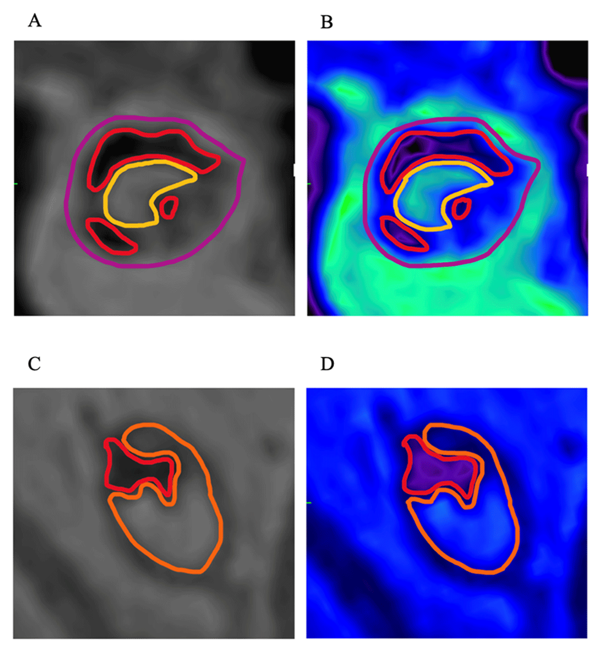
Figure 1
(A,B) Conventional greyscale and color-coded ultrashort echo time magnetic resonance imaging (UTE MRI) images of stenotic lesion; yellow outline: patent lumen; purple outline: thickened vessel wall with lipid rich and collagenous deposits typical in atherosclerosis; red outline: concentric and central calcium deposits. (C,D) Conventional greyscale and color-coded UTE MRI images of occlusive lesion; orange outline: soft collagenous component; red outline: eccentric calcium deposit.
This distinction is critical, as hard plaques have been found to markedly increase the risk of technical failure of percutaneous vascular intervention (PVI). This approach has demonstrated superior predictive value for PVI outcomes compared to conventional anatomical classifications such as the Trans-Atlantic Inter-Society Consensus (TASC) and Global Limb Anatomic Staging System (GLASS). It also offers the added benefits of being contrast-free and radiation-free, particularly advantageous in patients with renal insufficiency.41,42 MRA-defined “hard” lesions (> 50% lumen occlusion by calcium or dense collagen) had an 83% immediate technical failure rate, while “soft” lesions had only a 3% failure rate.40
With the incorporation of this cutting-edge imaging method into clinical workflows, our team aims to improve diagnostic sensitivity, individualize treatment regimens, and ultimately achieve higher limb salvage rates in patients with complex lower extremity arterial disease. UTE MRA can be accomplished without contrast administration, avoiding contrast-induced renal damage (not infrequent in PAD) and exposure to ionizing radiation, in contrast to CTA or X-ray angiography (Table 1).
Table 1
Summary of technical advantage of novel ultrashort echo time magnetic resonance imaging (UTE MRI) protocol. CTA: computed tomography angiography; NPV: negative predictive value
| FEATURE | UTE MRI | CONVENTIONAL MRI | CTA |
|---|---|---|---|
| Calcium and collagenous tissue | Clearly visualized | Signal void or artifacts | Blooming artifacts (especially calcium) |
| Soft/hard plaque distinction | Yes | Poor or none | Poor or none |
| Predictive value for PVI | High (sensitivity 83%, NPV 96%)21,22 | Low | Moderate |
| Radiation exposure | None | None | Present |
| Contrast requirement | Not needed | Often required | Required |
Integrating Unique MRA Protocol with Artificial Intelligence for Plaque Evaluation
Our research team has pioneered a novel and multidisciplinary approach for enhanced diagnostic accuracy for treatment planning in PAD by combining UTE MRI protocols with advanced advanced AI frameworks. A previously published foundational project of our research group is focusing on the development of a variational autoencoder (VAE)-based classification model utilizing 7T UTE MRA data obtained from amputated lower limbs from patients with PAD. By incorporating CNNs into the VAE framework, we trained the model on 2,390 pseudo-color reconstructed MRA images generated through multiplanar reconstruction and RGB fusion of UTE, T1-weighted, and T2-weighted sequences.
The AI algorithm thoroughly sorted different plaque regions into four categories according to luminal patency and tissue composition: (1) patent lumen, (2) partially patent with soft tissue, (3) predominantly occluded with soft tissue, and (4) predominantly occluded with hard tissue. This strategy established a semisupervised standardized scoring system for tissue assessment that directly maps to plaque type and severity from imaging data, thereby generating an interpretable latent space for vascular pathology studies. The findings demonstrated that the model effectively discriminated between soft and hard lesions and revealed the heterogeneity of the assessed lesions within the same patients, thereby showing its great potential to guide clinical decision-making.43
To further extend this methodology into an even higher resolution environment using 9.4T MRI scanners, our team introduced a study in which six vessel samples were scanned ex vivo, yielding a larger dataset of 4,014 cross-section images derived from multiplanar pseudo-color reconstruction. We analyzed this dataset using the previously introduced adapted VAE model with unsupervised clustering via a Gaussian mixture model in latent space. This time, the model autonomously identified four key lesion types—concentric calcified, eccentric calcified, hard occlusion, and soft occlusion. The VAE also assigned numerical tissue scores ranging from 0 (patent lumen) to 5 (hard occlusion) based on the proportion and spatial distribution of soft (eg, thrombus, fat) and hard (eg, calcification, dense collagen) tissue components. Most importantly, it achieved near-perfect classification accuracy for most classes and demonstrated 100% inter-rater agreement in predicting lesion crossability, defined as the ability to pass a guidewire through the occlusion during PVI. This ability to discern non-crossable, calcified, or fibrotic lesions from softer, guidewire-passable occlusions represents a substantial advancement in preprocedural planning and intervention risk stratification.44
Building on these successes, our most recent effort aimed to bring this AI-powered classification methodology closer to real clinical practice. Specifically, we tested the feasibility of using only UTE sequences acquired on a standard 3T clinical MRI scanner, thereby eliminating the need for long, multicontrast imaging protocols. Initially, a total of 797 arterial cross-sections from five amputated limbs of CLTI patients who underwent 3T scanning with a single UTE protocol were analyzed with our convolutional VaE model. The total scanning time in each case was under 15 minutes. Our early results already show the potential of the model to effectively distinguish soft and hard occlusions (Table 2). By significantly shortening acquisition time and avoiding the dependency on research-grade hardware or multisequence protocols, this method has great potential for clinical translation of our AI-assisted vascular imaging framework (Figure 2).
Table 2
Comparison of clinical 7T and preclinical 9.4T and clinical 3T magnetic resonance imaging (MRI)-based AI models for PAD lesion classification. AI: artificial intelligence; PAD: peripheral artery disease; VAE: variational autoencoder; MPR: multiplanar reconstructions; UTE: ultra-short echo time; CNN: convolutional neural network; RGB: red, green, blue
| FEATURE | VAE & 7T MRI STUDY | VAE & 9.4T MRI STUDY | VAE & 3T MRI STUDY |
|---|---|---|---|
| MRI field strength | 7 Tesla (clinical ultra-high field) | 9.4 Tesla (preclinical ultra-high field) | 3 Tesla (clinical standard field) |
| Sample size | 5 amputated limbs | 6 amputated limbs | 5 amputated limbs |
| Image dataset | 2,390 MPR pseudo-color images | 4,014 MPR pseudo-color images | 797 convention greyscale images |
| Input imaging sequences | T1w, T2w, UTE | T2w, UTE (RGB composite) | UTE only |
| AI model used | VAE with 2D CNN | VAE + Gaussian mixture model (GMM) in latent space | 2D convolutional VAE |
| Classification approach | Semi-supervised; manual quadrant division in latent space | Fully unsupervised; clustering via GMM in latent space | Unsupervised; tissue score via Euclidean distance in latent space |
| Output classes | (1) Patent lumen, (2) partially patent, (3) soft occlusion, (4) hard occlusion | (1) Concentric calcified, (2) eccentric calcified, (3) hard occlusion, (4) soft occlusion | Patent to occlusive continuum, differentiated by soft vs hard tissue features |
| Tissue scoring system | Numeric tissue score (TS: 0-5) for each lesion | Latent space clustering; classification probabilities | Tissue score based on latent space separation (0-1.26) |
| Key strength | Clinically feasible, interpretable results, real-world applicability | High resolution, high diagnostic precision, excellent inter-rater reliability | Short acquisition time, no contrast or multi-sequence required, high scalability |
| Crossability prediction | Inferred through TS and plaque type | Achieved 100% inter-rater reliability in predicting lesion crossability | Aligned with expert visual assessment; scalable clinical proxy for lesion severity |
| Clinical integration potential | High–faster scans, easier implementation | Lower–limited by cost, availability, and scan time | Very high–compatible with clinical MRI workflows, < 15 min scan time |
| Best use case | Real-time clinical support and triage | Advanced preprocedural planning and research use | Point-of-care decision-making; standard clinical PAD assessment |
| Main contribution | Introduced AI-based semi-automated TS classification for PAD | Demonstrated unsupervised deep learning for lesion phenotyping with guidewire relevance | Validation of feasibility of using only UTE sequence on 3T clinical scanner for lesion assessment |
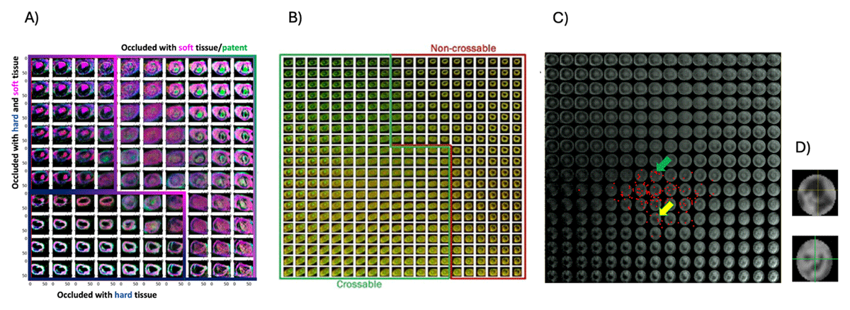
Figure 2
(A) Peripheral artery disease tissue type groups in the latent space separated by a custom-made variational autoencoder from magnetic resonance imaging-histology images.43 (B) Data in latent space generated by the variational encoder showing crossable and non-crossable lesions. Hard tissues are represented by dark green/black colors, whereas soft tissues are represented by lighter red/green colors.44 (C) Depiction 2D latent space illustrating the separation of the patent lumen (upper right) to occlusion (lower left). Red dots indicate the location of cross-sections in (D) calculated by the decoder of the convolutional variational autoencoder. Colored arrows point to the approximate location of lesions in (D); yellow: occlusive; green: partially patent
Collectively, these AI-powered frameworks highlight the tremendous transformational potential of integrating UTE MRI with advanced deep learning tools to improve the diagnostic precision and clinical utility of PAD imaging. The 7T VAE-based model illustrates the feasibility for fast, semiautomated vascular lesion classification in a clinically accessible setting. By contrast, the 9.4T-based crossability study illustrates the utility of ultra-high-resolution imaging for the identification of technically challenging, non-crossable lesions with high inter-rater agreement. By continuing to refine these platforms, we aim to close the diagnostic gap between imaging appearance and histopathological reality, ultimately enabling personalized, predictive, and more effective care for patients with complex peripheral artery disease.
Summary
AI-based innovations can reshape the common practice of PAD care by enabling early diagnosis, personalized treatment, and efficient clinical workflows. While the current research landscape seems to favor CTA and ultrasound based on data availability and more standardized imaging protocols, MRA—especially the noncontrast scans—has significant potential due to its superior tissue characterization and resolution. If combined with AI-based analysis, MRA has the potential to become a powerful modality for PAD detection and treatment planning and to translate disease management into more accessible, timely, and personalized care. Future research should prioritize prospective validation, interoperability, and human-centered design to translate AI innovations into daily vascular practice.
Key Points
Review of existing studies shows opportunities and challenges for clinical translation of deep learning-based methods in vascular care.
Our research group introduces a novel magnetic resonance imaging (MRI) protocol for vascular lesion characterization in peripheral artery disease.
Artificial intelligence combined with MRI can enhance noninvasive assessment of vascular plaque composition.
Accurate lesion characterization supports improved treatment planning in peripheral artery disease.
Competing Interests
The authors have no competing interests to declare.