
A 58-year-old male with a history of hypertension and atrial fibrillation presented with substernal chest pain and sinus tachycardia. He had no previous history of cardiovascular disease. A CT angiogram was performed to rule out pulmonary embolism, which incidentally revealed a right-sided aortic arch with an aberrant left subclavian artery and associated Kommerell’s diverticulum as shown in Figure 1 and 2. The diverticulum was causing mild tracheal and esophageal compression. The patient denied symptoms such as dysphagia or odynophagia. Transthoracic echocardiography revealed no valvular abnormalities. Coronary imaging and stress testing were unremarkable. Although surgical follow-up was recommended due to the anatomic findings, the patient left against medical advice.
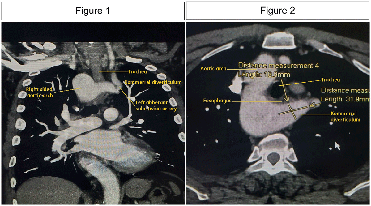
Figures 1 and 2
Axial and coronal maximum intensity projection (MIP) images of contrast-enhanced computed tomography (CECT) demonstrate a right-sided aortic arch with an anomalous origin of the left subclavian artery. There is evidence of saccular dilatation at its origin, consistent with Kommerell’s diverticulum.
Ethics and Consent
Written informed consent was obtained from the patient for the publication of this case report. The patient was shown the manuscript and accompanying images and consented to their publication.
Competing Interests
The authors have no competing interests to declare.