
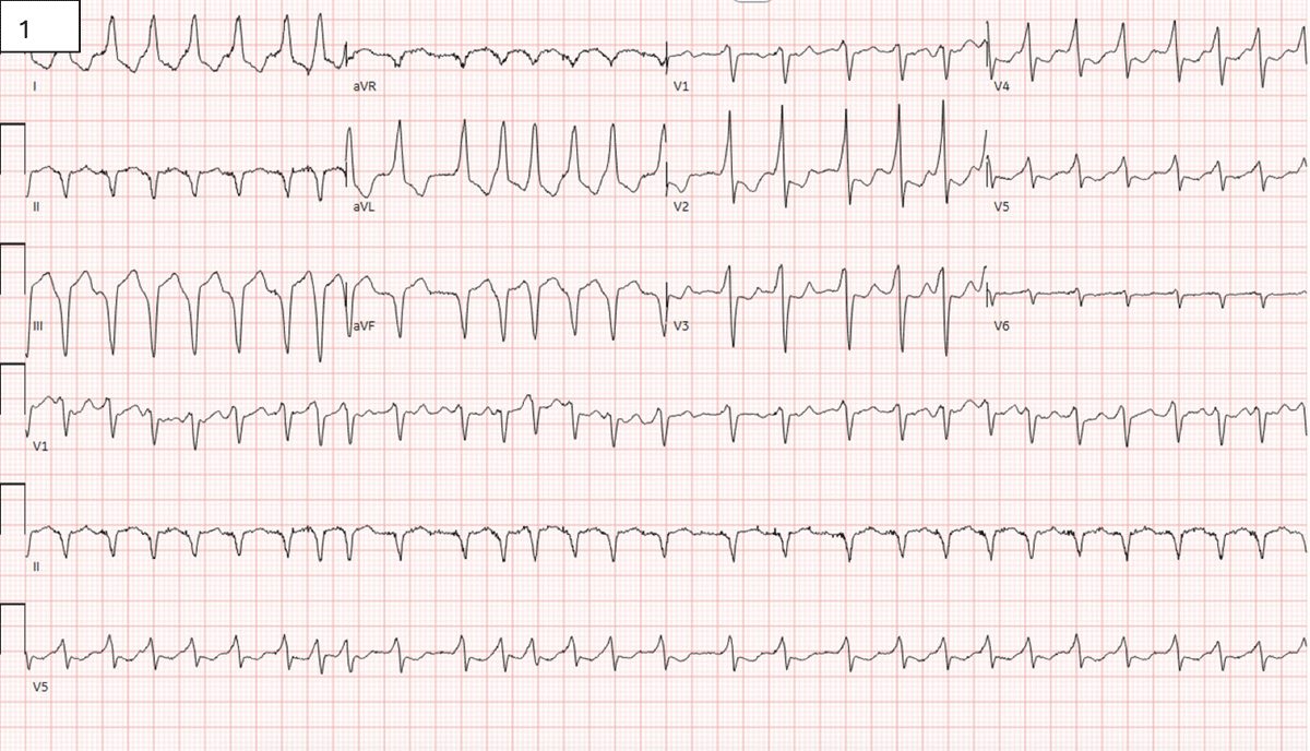
Figure 1
Electrocardiogram demonstrating atrial fibrillation with rapid ventricular rate, nonspecific intraventricular conduction block, and marked ST segment changes concerning for anterior subendocardial injury. There is also ST segment depression in lateral leads and Q waves in inferior leads.
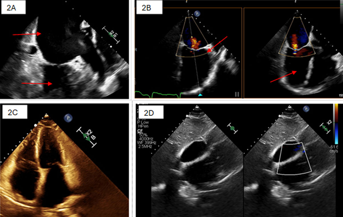
Figure 2
(A,B) Transesophageal echocardiogram demonstrates severe dilation of the left and right atria with no thrombus. The left ventricle (LV) exhibits significantly reduced systolic function (ejection fraction 20-25%), with global hypokinesis; the right ventricle shows mild to moderate systolic dysfunction. Mild mitral and moderate tricuspid regurgitation are present (2B), with central jets in both valves. (C,D) Transthoracic echocardiogram demonstrates persistent decreased systolic function and low ejection fraction of LV after cardioversion.
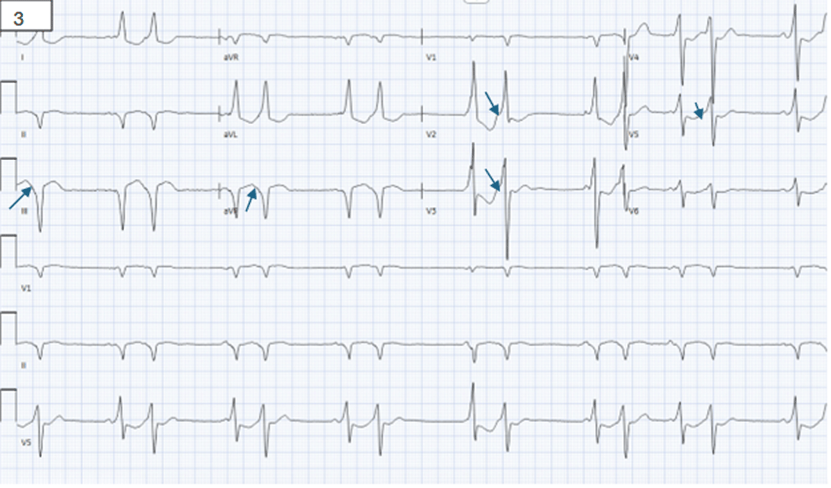
Figure 3
Electrocardiogram demonstrating normal sinus rhythm with preexcitation pattern, negative delta waves in the inferior leads, and positive delta waves in the anterior leads as depicted by the arrows.
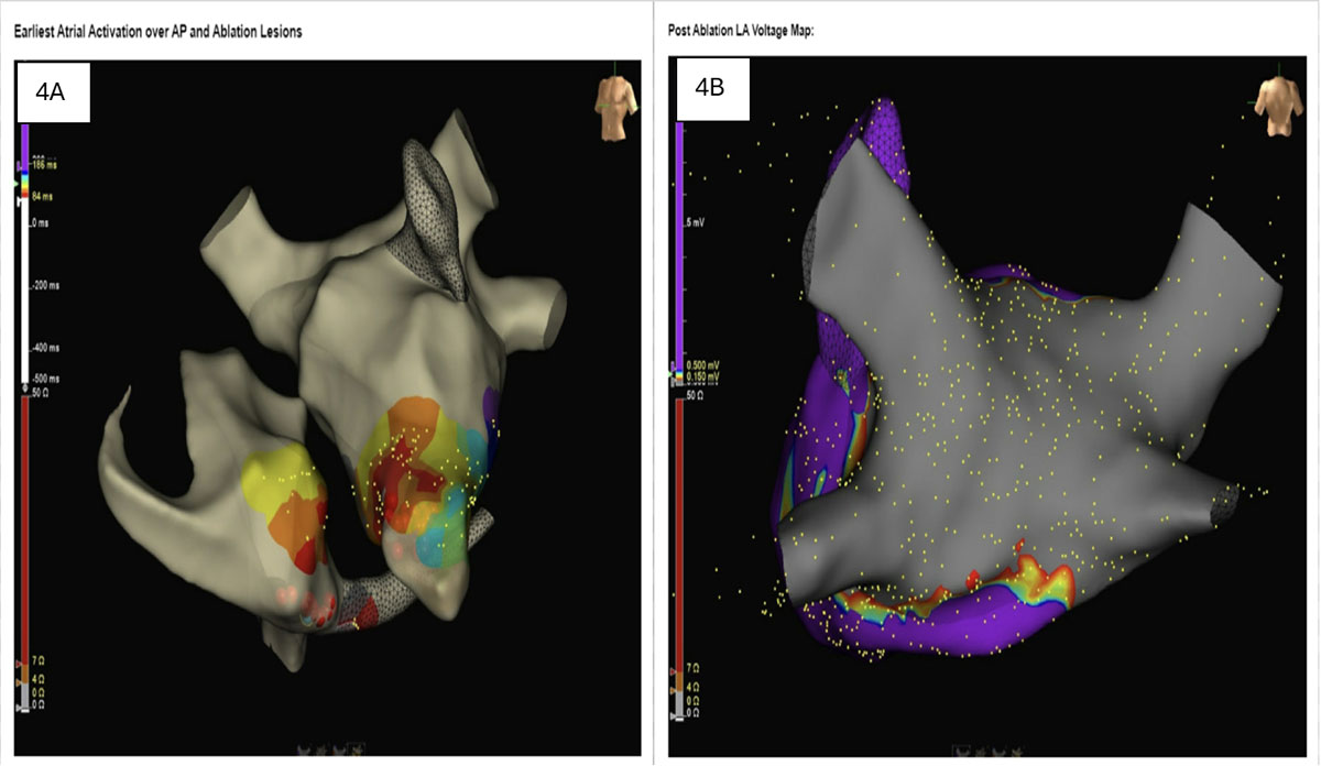
Figure 4
(A) Mapping illustrating the earliest atrial activation and potential accessory pathway. (B) Successful ablation of the accessory pathway in the right posteroseptum, with no remaining evidence of the pathway.
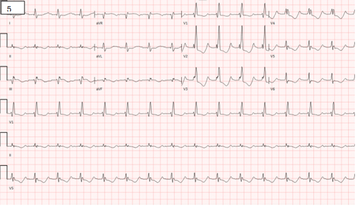
Figure 5
Electrocardiogram demonstrates sinus rhythm with first-degree atrioventricular block, right atrial enlargement, and right bundle branch block. Preexcitation is no longer observed.
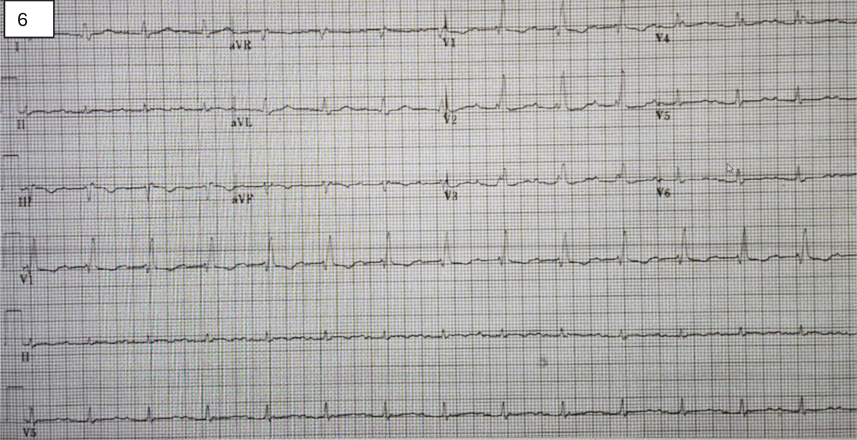
Figure 6
Electrocardiogram at 2 months showed normal sinus rhythm with no recurrence of atrial fibrillation.