
Introduction
Anomalous coronary arteries are rare congenital heart defects occurring in less than 1% of the general population but are increasingly recognized with the growing use of chest imaging, specifically computed tomography (CT) and magnetic resonance imaging (MRI). They fall into two main categories: abnormal coronary artery origin and anomalous vessel development, such as fistulae or aneurysms. Some occur in conjunction with other congenital heart defects (eg, transposition of the great arteries, truncus arteriosus, or tetralogy of Fallot), while others appear in isolation. Proper risk stratification is essential, as many patients are asymptomatic while others face complications such as myocardial infarction, ventricular arrhythmias, or sudden cardiac death. This review explores the three major types of anomalies, including anomalous origins, aneurysms, and fistulae (Table 1).
Table 1
Summary of prevalence, evaluation methods, and treatment for anomalous coronary arteries. R-AAOCA: right anomalous aortic origin of coronary artery; L-AAOCA: left anomalous aortic origin of coronary artery: ARCAPA: anomalous right coronary artery from the pulmonary artery; ALCAPA: anomalous left coronary artery from the pulmonary artery; CAF: coronary artery fistula; CCTA: coronary computed tomography angiography; CABG: coronary artery bypass graft; PCI: percutaneous coronary intervention
| ANOMALY | PREVALENCE (PER 100,000) | REPAIR INDICATED? | IDEAL IMAGING MODALITY | SIGNIFICANT FEATURES TO EVALUATE | TREATMENT OPTIONS |
|---|---|---|---|---|---|
| R-AAOCA | 0.33% (330)3 | Symptoms or ischemia | CCTA | Interarterial course, intramural segment, slit-like ostium, AAOCA dominance | Unroofing, reimplantation |
| L-AAOCA | 0.12% (120)3 | Symptoms or ischemia | |||
| ARCAPA | 0.002% (2)22 | Yes | Collaterals, dominance | Reimplantation, ligation, CABG, channel surgery | |
| ALCAPA | 0.008% (8)22 | Yes | |||
| CAF | 0.002% (2)33 | Symptoms | Fistula course, size, collaterals | Percutaneous intervention, surgical ligation +/- bypass | |
| Aneurysm | 0.3-5.3% (300-5,300)48,49 | Symptoms | Calcification, luminal diameter, thrombi and aneurysmal features | PCI, CABG +/- ligation |
Abnormal Coronary Origin
Anomalous Aortic Origin
Anomalous aortic origins of the coronary arteries (AAOCA) vary greatly in anatomy, patient presentation, and optimal treatment. The prevalence of AAOCA in the general population is reported to be 0.2% to 0.5% based on several autopsy studies.1,2 The most common and clinically significant form of anomalous aortic origin is coronary arteries originating from the opposite sinus of Valsalva. In a cardiac MRI screening study of 5,169 healthy children, AAOCA from the opposite sinus of Valsalva had a prevalence of 0.44%, with 0.33% being right coronary arteries from the left sinus (R-AAOCA) and 0.12% being left coronary arteries from the right sinus (L-AAOCA).3
AAOCA anatomical variations directly influence ischemic risk and management decisions. Coronary arteries may originate from the aorta via separate, shared, or branch ostia and are classified based on their course as interarterial, subpulmonic (intraconal/intraseptal), pre-pulmonic, retroaortic, or retrocardiac.4 Interarterial vessels travel between the aorta and the pulmonary artery (PA) and are generally considered the most high-risk. Ischemic risk, while previously attributed primarily to vessel compression between the aorta and PA, is now associated with additional features, including a slit-like (fish-mouth) orifice, acute take-off angle, proximal vessel hypoplasia, and particularly an elongated intramural segment.4 A slit-like orifice combined with an intramural segment can create valve-like obstruction, significantly restricting coronary flow and heightening the risk of sudden cardiac death (SCD).5
Clinical presentations vary considerably, from asymptomatic incidental discovery to cardiac symptoms such as chest pain, dyspnea, syncope, or even SCD.6,7,8 SCD predominantly affects pediatric patients and young adults involved in competitive sports, with significantly lower incidence beyond age 35,9 and many do not recall prior anginal symptoms.10,11,12 These patterns underscore the need for careful anatomical and physiological evaluation for effective risk stratification.
Imaging plays a central role in both the anatomical characterization and functional assessment of anomalous coronary arteries, guiding clinical decision-making between surgical and conservative management.5 Coronary computed tomography angiography (CCTA) is the gold standard for delineating proximal coronary anatomy, reliably identifying high-risk features such as intramural course, slit-like ostium, and acute take-off angle (Figure 1). Invasive angiography can also be helpful in evaluating ostial characteristics under dobutamine infusion by intravascular ultrasound and flow assessment.13,14
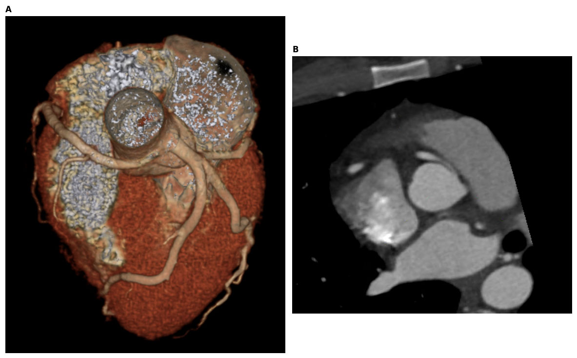
Figure 1
A 33-year-old man with R-AAOCA diagnosed as an adult. (A) CCTA shows the anomalous origin of the dominant RCA from the left aortic sinus of Valsalva, (B) with slit-like ostium and intramural course. R-AAOCA: right anomalous origin of the left coronary artery; CCTA: coronary computed tomogram angiography; RCA: right coronary artery
Cardiac MRI complements CCTA by providing radiation-free evaluation of myocardial perfusion and scar, making it particularly valuable in younger patients. Functional testing—especially with dobutamine stress protocols using echocardiography, cardiac MRI, or positron emission tomography—is essential for detecting dynamic components of ischemia that may be missed by vasodilator-based or submaximal stress tests. The addition of a dobutamine-volume challenge helps mimic exertional conditions and can unmask phasic compression in the intramural segment.15
Recent advances in computational modeling, including patient-specific 3D printing and computational fluid dynamics (CFD), have enhanced surgical planning by enabling more accurate predictions of hemodynamic consequences and procedural risks. CFD simulations, which assess flow dynamics and wall shear stress in AAOCA, show promise in improving risk stratification and guiding intervention strategies.16,17,18 However, current methodologies remain heterogeneous, and standardization is necessary to validate and incorporate these techniques reliably into routine clinical practice.
Surgical intervention for AAOCA is recommended for symptomatic patients, those with documented coronary ischemia, or asymptomatic individuals with high-risk anatomical features.5 However, an increasing number of patients diagnosed incidentally later in life may be safely managed medically after thorough evaluation.14 The most common surgical approach is unroofing of the intramural coronary segment, a procedure with high success rates and minimal operative risk.19,20,21,22 Alternative surgical techniques include coronary reimplantation (which requires mobilization of a longer segment of the artery) and, rarely, coronary artery bypass grafting (generally reserved for concomitant atherosclerotic disease because the intermittent coronary flow reduction associated with AAOCA is usually not enough to promote maturation of a venous or arterial graft).
Percutaneous coronary stenting also has been reported, typically used to treat comorbid coronary artery atherosclerosis, though there may be an emerging role to treat the ostial segment itself as radial strength and coronary stent technology improves.23,24 This method should generally be limited to nonsurgical candidates due to insufficient long-term efficacy data.
Anomalous Origin from Pulmonary Artery
Anomalous origin of a coronary artery from the PA is another important coronary anomaly that presents distinct clinical challenges. The two primary variants are anomalous right coronary artery from the PA (ARCAPA) and anomalous left coronary artery from the PA (ALCAPA), also known as part of the Bland-White-Garland syndrome.25 Though less common than AAOCA, these anomalies more frequently cause symptomatic ischemia. Their combined incidence in the general population is approximately 0.01%.25,26 Among patients undergoing coronary angiography, ALCAPA (0.008%) is more common than ARCAPA (0.002%), likely due to anatomical proximity between the left coronary artery and the PA.25
The clinical presentation of ALCAPA and ARCAPA largely depends on coronary artery dominance and collateral circulation (Figure 2A). When a dominant coronary artery lacks sufficient collaterals, myocardial ischemia can occur, particularly during the neonatal period when pulmonary vascular resistance is high.27 Additionally, the lower diastolic pressure of the anomalous origin from the PA will not provide adequate perfusion pressure. As pulmonary pressures decrease, blood may flow retrogradely from the coronary artery into the PA, known as the “coronary steal” phenomenon, further exacerbating ischemic injury.28
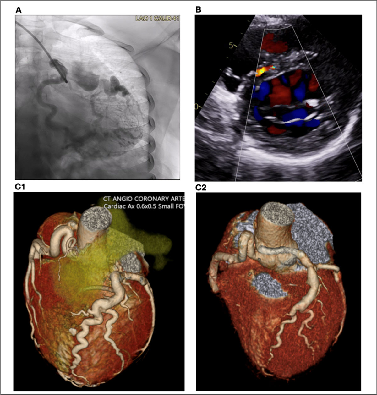
Figure 2
A 26-year-old woman with ALCAPA diagnosed as an adult. (A) Preoperative coronary angiogram shows a large RCA with septal and epicardial collaterals to the LAD and LCX that converge into LMCA draining into the PA. (B) Preoperative transthoracic echocardiography with color Doppler reveals collateral vessels in the interventricular septum. (C1,2) Pre- and postoperative CCTA demonstrate the abnormal origin of LM from the PA and postsurgical SVG graft placement, respectively. ALCAPA: anomalous left coronary artery from the pulmonary artery; RCA: right coronary artery; LAD: left anterior descending artery; LCX: left circumflex artery; LMCA: left main coronary artery; PA: pulmonary artery; cCTA: coronary computed tomogram angiography; SVG: saphenous vein graft
Patients typically present either as infants or in late adulthood, and patients with ALCAPA are more likely to be symptomatic/diagnosed earlier than patients with ARCAPA.28,29 Patients without sufficient collaterals typically present as infants with symptoms such as profuse sweating, dyspnea, pallor, and atypical chest pain while eating or crying. If left untreated, symptomatic infant patients have a mortality rate up to 90%.30 Patients who have established collateral vessels are mostly asymptomatic or may present later in life with symptoms such as mitral insufficiency, ischemic cardiomyopathy, or malignant dysrhythmias.28 Even asymptomatic adult patients have an increased risk of poor outcomes including sudden cardiac death.31 The American Heart Association and European Society of Cardiology Guidelines for Adult Congenital Heart Disease recommend surgery for all patients with ALCAPA and symptomatic patients with ARCAPA (Class I recommendations).5,32
CT angiography is the preferred imaging modality for ALCAPA and ARCAPA (Figure 2C).28,33 Yet among the coronary anomalies discussed, echocardiography is particularly informative in the evaluation of ALCAPA. Key findings include the absence of a left coronary origin from the aorta and direct visualization of its anomalous connection to the PA. A markedly dilated right coronary artery is often seen due to increased collateral flow (Figure 2B). Color Doppler imaging can demonstrate retrograde flow from the left coronary artery into the PA and visualize septal collateral vessels. Additional findings typically include left ventricular dilation and systolic dysfunction as well as mitral regurgitation secondary to ventricular dilation or papillary muscle ischemia. Together, these echocardiographic features are critical for the early diagnosis and appropriate management of ALCAPA.34,35
Re-implantation, which may involve a transpulmonary baffle (Takeuchi procedure), is the preferred surgical approach when feasible as it restores the two-vessel coronary artery system originating from the aorta and corrects the coronary steal phenomenon.28,33 Other possible operations include ligation of the coronary artery and bypass grafting, although graft patency is suboptimal in adults because the anomalous coronary is large and requires high flow to compete against the well-developed contralateral collaterals.36
Exercise Restriction & Long-Term Follow-Up of AAOCA
Recommendations on exercise restriction in patients with anomalous coronaries vary based on the anomalous origin and the identification of high-risk anatomical features. Per the AHA Guidelines on Clinical Considerations for Competitive Sports Participation for Athletes with Cardiovascular Abnormalities, L-AAOCA with interarterial course should generally undergo surgical correction prior to strenuous exercise, and competitive sports should be avoided if the anomaly is left unrepaired. R-AAOCA with interatrial course is considered lower risk and, when unrepaired, is not a contraindication to exercise if evaluation for exertional cardiac symptoms and/or inducible ischemia is negative.37,38
Patients with ALPACA who have undergone repair in infancy can return to competitive sports after surgical healing provided that LV function is normal and ischemia cannot be induced. Patients with lower-risk coronary anomalies that do not involve an interarterial course or arise from the PA can generally participate in strenuous exercise unless there is associated evidence of ischemia. Exercise restrictions can be distressing, especially for young patients, and may be associated with adverse long-term cardiovascular outcomes. In all cases, shared decision-making should be practiced and should include individualized risk counseling based on an assessment of the patient’s specific anatomy.37,38
Patients with anomalous coronaries who have undergone surgical or percutaneous intervention should be followed longitudinally, ideally by a multidisciplinary heart team at a center of expertise.5 It is plausible that anomalous coronaries may be predisposed to coronary artery disease (CAD) due to tortuous routes and turbulent blood flow within the vessel, thus warranting more intensive lifelong primary prevention with strict lipid and blood pressure control, although data on this are limited.39,40 There are no guidelines on specific primary atherosclerotic cardiovascular disease prevention strategies for patients with anomalous coronaries, among other CAD.
In women of childbearing age, pregnancy may be a period of increased risk due to hemodynamic changes, hormonal changes, and potential aortic dilation. In one study including women with coronary artery anomalies secondary to CAD or coronary aneurysm secondary to inflammatory disease, there was a 14% risk of major adverse cardiovascular event during pregnancy.41 Patient registries can help to guide further research on perioperative and long-term outcomes for patients with anomalous coronaries.42
Incomplete or Abnormal Development
Fistulae
Coronary artery fistulae (CAF) involve abnormal communication between at least one coronary artery and either of the cardiac chambers or the great vessels adjacent to the heart. While CAF can be acquired, 90% are congenital—although congenital CAF are rare, with a reported incidence of 0.002% in the general population.43,44 The incidence of CAF in patients undergoing coronary angiography is reported to be higher, between 0.1% and 0.9%.46,47,48 The most common origin of CAF is from the LAD, and they tend to drain into the PA, left ventricle, and right heart.49,50,51
CAF can cause myocardial ischemia primarily through three mechanisms: (1) diversion of blood flow from coronary branches into the fistula tract; (2) thrombus formation in the dilated coronary and downstream embolization; or, more rarely, (3) stenosis of coronary side branches secondary to fistula tracts, ulcerations, and atherosclerosis.52 While most CAF are asymptomatic and discovered incidentally during cardiac catheterization, symptomatic patients may present with chest pain, exertional dyspnea, or palpitations, which may be suggestive of underlying arrhythmias or myocardial ischemia.32,45 Larger or more complex fistulae can lead to severe heart failure or pulmonary congestion.47 Upon diagnosis of a CAF, CT angiography is essential to characterize its anatomical pathway and features, guiding subsequent management decisions.47
Most asymptomatic patients with small fistulae may be safely monitored without treatment.47,53 For symptomatic patients, current European and American guidelines recommend closure of larger CAF, either by surgical or percutaneous intervention (Class I recommendation).5,32 Percutaneous intervention is appropriate for patients with a narrow single CAF who have no other associated cardiac anomalies.47,54 Conversely, surgical repair is indicated for aneurysmal, tortuous, or ectatic CAF and typically involves simple ligation of the fistula, although hybrid surgical-transcatheter approaches may be beneficial for complex anatomy.51,54,55 Notably, surgical treatment carries significant perioperative risks, including myocardial infarction (11%) and late mortality (24%).56
Aneurysms
Coronary artery aneurysms (CAAs) are defined as a coronary artery segment that is dilated 1.5 times more than the diameter of the adjacent segment.57 The incidence of CAAs varies from 0.3% to 5.3%.58,59,60 Coronary artery aneurysms can be morphologically classified as either saccular or fusiform, involving the tunica layers of the vessel wall, while its dilatation may be focal or diffuse.61,62 If the dilation is more than four times the surrounding vessel or greater than 8 mm in diameter, it is known as a giant aneurysm.62 CAAs are predominantly found in men and commonly observed in the right coronary artery.57,61
CAAs can be congenital, acquired, or iatrogenic. Congenital aneurysms account for 20% to 30% of all CAAs and may result from genetic disorders—such as connective tissue diseases like Marfan syndrome—or from fistulous communication and turbulent blood flow between a coronary artery and another vessel or cardiac chamber.62,63 Atherosclerosis is a leading cause of acquired CAA, occurring in 50% of adult cases.61 In children, inflammatory conditions including Kawasaki disease and multisystem inflammatory syndrome in children (MIS-C) are a common cause of acquired CAAs.64,65,66 In Kawasaki disease, vascular inflammation occurs with neutrophilic infiltration leading to endothelial destruction in the vessel wall and aneurysm formation. Subsequent lymphocytic infiltration can also lead to subacute vasculitis. Inflammation may also lead to thrombus formation with resulting dilation of the proximal blood vessel.64,66
MIS-C is a life-threatening complication following coronavirus disease 2019 (COVID-19) infection in children. CAAs are estimated to occur in 8% to 24% of patients with MIS-C as a result of epicardial inflammation.65 Additional infectious causes (eg, syphilis, Lyme, mycotic aneurysms secondary to bacterial or fungal endocarditis) can cause immune complex deposition, vessel wall disruption, and aneurysm formation. Iatrogenic aneurysm can arise from vessel injury during percutaneous interventions, including stenting and atherectomy.67
Molecularly, aneurysm formation involves the breakdown of extracellular matrix structures due to increased matrix metalloproteinase (MMP-2 and MMP-9) activity coupled with inflammation driven by cytokines (IL-1β, IL-6, TNF-α) and altered vascular remodeling mediated by growth factors such as TGF-β and PDGF.68 Targeting these molecular pathways could offer new therapeutic strategies to halt aneurysm progression and prevent associated cardiovascular complications.
Depending on what the aneurysmal wall is composed of, CAAs can be classified as either true or false aneurysms (pseudoaneurysm).69 True aneurysms are characterized by normal vascular integrity, while pseudoaneurysms are formed of extravascular hematomas that communicate with intravascular space and are formed with thin-walled structures that don’t have normal arterial wall layers.69
Like CAF, CAA is an uncommon entity that is often recognized incidentally by coronary angiography, computed tomography angiography, or autopsy.58,67 In terms of invasive imaging modalities, coronary angiography is the gold standard for the evaluation of CAAs. Furthermore, intravascular ultrasound is also highly valuable as it provides detailed information about the arterial wall, differentiates true aneurysm from pseudoaneurysm, and assesses the effects of plaque rupture on the aneurysms.58 CAAs are evaluated by identifying their characteristics, such as their maximal diameter, distribution, presence or absence of intraluminal thrombi, the number and extension of the aneurysms, and potential complications such as myocardial infarction.60
CAAs are mostly asymptomatic, but patients who do have symptoms typically present with effort angina.58 They can lead to complications such as ischemia, vessel thrombosis, fistula formation or rupture, and sudden cardiac death or congestive heart failure.58,70,71 Most of these complications can be associated with the turbulent flow linked to the aneurysms, although they could be characteristics of atherosclerotic coronary artery disease.62
Similar to other coronary artery anomalies, CAAs can be treated by various methods that include medical treatment, percutaneous coronary intervention (PCI) such as stent angioplasty, and surgical excision.59,72 PCI is preferred to remove smaller CAAs (< 10 mm in diameter) whereas a surgical approach is relevant for larger aneurysms.62,67 Bypass grafting has been extensively studied as the treatment approach.62 In cases where the aneurysm is three times larger than the original vessel’s diameter, some surgeons suggest mandatory surgical intervention.69 Based on anecdotal evidence, CAAs are medically managed by using antithrombotic/anticoagulant and antiplatelet agents.69 The success rates of the treatments mentioned above are unclear due to the lack of enough clinical trials, and treatment of CAAs has been particularly challenging due to the lack of standardized international recommendations. Therefore, treatment of CAAs is mostly individualized and relies on factors such as clinical presentation, characteristics, the patient’s profile, and the physician’s experience.67
As alluded to earlier in this review, CFD has been increasingly utilized for risk stratification in AAOCA. By using patient-specific imaging, CFD simulations can provide detailed hemodynamic information such as flow patterns and wall shear stress, crucial for predicting thrombotic risks.73,74,75 While CFD enables individualized assessments beyond traditional aneurysm measurements, its accuracy depends on imaging quality, modeling assumptions, and computational demands, potentially limiting routine clinical use. Nonetheless, integrating CFD into clinical practice holds promise for improved CAA management.
Conclusion
Coronary artery anomalies vary widely in their morphology, presentation, and clinical risk to patients. Many asymptomatic patients with anomalous coronary arteries identified incidentally in adulthood may be managed conservatively. Whether a patient requires surgical or percutaneous repair of an anomalous coronary artery depends on the patient’s symptoms, the course/features of the artery, evidence of ischemia, patient preferences on degree of desired activity and risk tolerance, and the judgement of the clinical team. Consequently, the course and morphology of anomalous coronary arteries must be thoroughly examined to guide treatment decisions, and CT angiography is the gold standard for doing so. Further research is required to determine the necessity (or lack thereof) of treating certain coronary anomalies, such as in certain forms of AAOCA.
Key Points
Coronary artery anomalies are a rare congenital condition that are being increasingly identified due to better imaging modalities.
The severity of risk of anomalous coronary arteries depends on multiple characteristics such as their course, size, narrowing, dominance, and collaterals.
Management of patients with anomalous coronary arteries depends largely on their symptoms, signs of ischemia, and artery characteristics.
Repair of anomalous coronary arteries includes surgical repair, such as unroofing, as well as percutaneous methods.
Competing Interests
Dr. Ada Stefanescu Schmidt is a consultant for reCross Cardio and B. Braun Medical Inc., and her research is partly supported by Edwards Lifesciences and Occlutech. The other authors have no competing interests to declare.