
A 75-year-old female with a past medical history of idiopathic small-fiber neuropathy, essential hypertension, and heterozygous familial hypercholesterolemia was evaluated by her cardiologist for fatigue and shortness of breath on exertion.
Electrocardiography showed normal sinus rhythm with first-degree atrioventricular block. To assess for possible ischemic etiology, the patient underwent coronary computed tomography angiography (CTA), which showed a left-dominant coronary system without significant stenosis but with mild atherosclerosis. Notably, a 16-mm wide-neck Windsock aneurysm of the interventricular septum was identified, along with a patent foramen ovale and left-to-right shunting (Figures 1, 2).
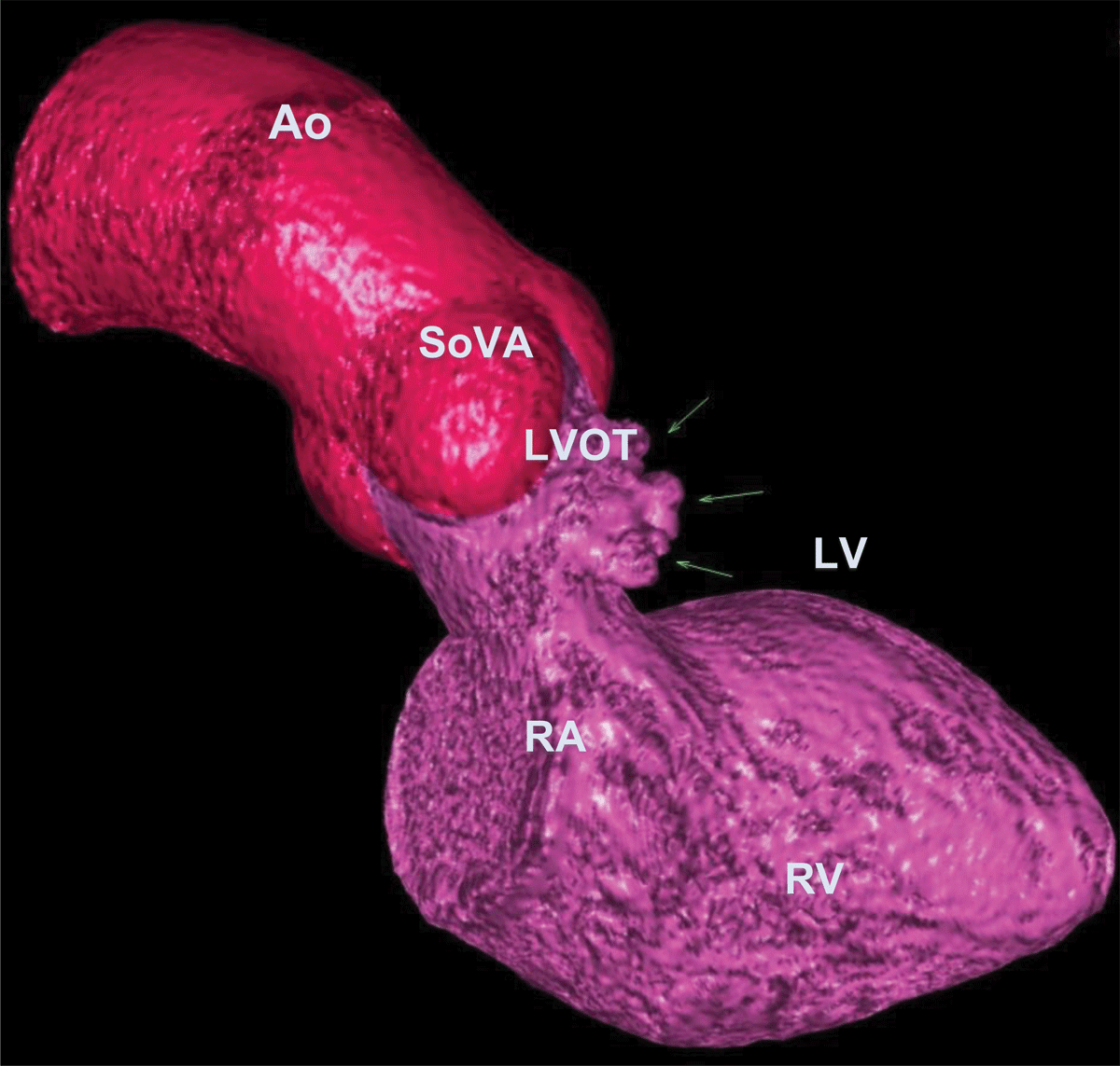
Figure 1
3D cardiac computed tomography angiography showing interventricular septal aneurysm (arrows). Ao: aorto; SoVA: sinuses of Valsalva; LVOT: left ventricular outflow trac; RV: right ventricle; LV: left ventricle; RA: right atrium
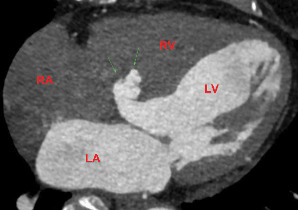
Figure 2
Cardiac computed tomography angiography showing interventricular septal aneurysm (arrows). LA: left atrium; LV: left ventricle; RA: right atrium; RV: right ventricle
Membranous interventricular septal aneurysms are rare congenital anomalies often discovered incidentally during evaluation for unrelated symptoms.1 Our patient underwent CTA for ischemic evaluation and was found to have an aneurysm. It is important to assess for associated structural abnormalities, such as sinus of Valsalva aneurysms, ventricular septal defects, or outflow tract anomalies such as transposition of the great arteries.2,3,4
While typically asymptomatic, these aneurysms can be a nidus for thrombus formation, increase the risk of arrhythmias, or cause right ventricular outflow tract obstruction if they protrude significantly.1,5 Although our patient was symptomatic, her symptoms were deemed unrelated to the aneurysm, and she did not have a thrombus or evidence of arrhythmia. Transthoracic echocardiography can aid in detection, but cardiac computed tomography imaging provides superior anatomical detail, allowing for better visualization and assessment of associated cardiac pathology.
Given the absence of significant coronary artery disease, the patient was managed conservatively with risk factor optimization for atherosclerosis and close follow-up for potential embolic risk assessment.
Competing Interests
The authors have no competing interests to declare.