
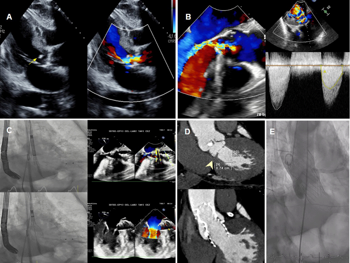
Figure 1
(A) Transthoracic echocardiography parasternal long-axis view showing subaortic membrane (arrow) with flow acceleration in the left ventricular (LV) outflow tract and below the aortic valve. (B) Transesophageal echocardiography (TEE) long-axis view showing subaortic membrane with flow acceleration in the LV outflow tract and below the aortic valve, continuous-wave Doppler showing a double-envelope pattern. The inner envelope on the right side of the image (yellow) represents stenosis at the subaortic membrane, while the outer envelope on the left side of the image (white) represents stenosis at the aortic valve. (C) A 26-mm Evolut™ PRO+ transcatheter aortic valve implantation (TAVI) was deployed with TEE guidance to position the valve precisely at the subvalvular ridge. (D) Cardiac computed tomography (CT) multiplanar reconstruction shows a subaortic membrane (arrowhead), and a 1-year post-TAVI CT demonstrates a well-seated valve covering most of the left ventricular outflow tract (LVOT), including the previously identified subaortic membrane. (E) Final aortography shows a well-seated valve in the LVOT covering the subaortic membrane, with no paravalvular leak.
Video 1
Transesophageal echocardiography showing subaortic membrane with flow acceleration in the left ventricular outflow tract and below the aortic valve; also see at https://youtube.com/shorts/-xcWHpuayeA.
Video 2
Aortography showing a well-seated valve in the left ventricular outflow tract covering subaortic membrane with no paravalvular leak; also see at https://youtube.com/shorts/kMOCuulhQ8I.
Video 3
Transthoracic echocardiography post transcatheter aortic valve implantation; also see at https://youtube.com/shorts/C-q8TdWPsW4.