
Introduction
Endomyocardial fibrosis is an uncommon condition, with the majority of cases traced to Brazil, Africa, and other tropical and subtropical countries.1 It is often characterized by fibrosis of the endocardium and myocardium, including the inlet and apex of either the right, left, or both ventricles. Most of the time, its clinical evolution is progressive, with findings of refractory heart failure.2 Idiopathic calcific cardiomyopathy (ICC) is a rare and poorly understood cardiac condition characterized by diffuse endomyocardial calcifications in the absence of an identifiable systemic disorder.3 This case report presents a 76-year-old female patient with a history of hypertension and hyperlipidemia who was initially evaluated for symptoms suggestive of coronary artery disease (CAD) but was ultimately diagnosed with ICC.
Case Presentation
A 76-year-old female from Texas presented with an 8-week history of exertional chest pain radiating to the left arm and alleviated by rest. Her medical history was significant for hypertension and hyperlipidemia and was managed with amlodipine besylate, benazepril, and rosuvastatin. The patient denied any family history of cerebrovascular accidents, myocardial infarction, or other significant cardiovascular events, and she reported no travel history to tropical regions.
On examination, the patient appeared comfortable and in no acute distress. Her pulse was regular at 74 beats per minute with normal wave and character. Blood pressure was 140/80 mm Hg. She was afebrile with a respiratory rate of 14 breaths per min and oxygen saturation of 96% at room air. Cardiovascular examination revealed a regular pulse with normal wave contour and character; auscultation revealed normal S1 and S2 heart sounds along with an S4 gallop suggestive of reduced left ventricular (LV) compliance, often seen in conditions such as hypertension or ischemic heart disease.
The initial diagnostic evaluation included a 12-lead electrocardiogram (ECG), which highlighted a normal sinus rhythm with poor R-wave progression in anterior leads and no evidence of acute infarction or ischemic changes. Given the patient’s presenting symptoms and initial ECG findings, further cardiac imaging was pursued to evaluate for myocardial ischemia and structural abnormalities. A Rubidium-82 positron emission tomography (PET) myocardial perfusion study was performed, revealing new reversible perfusion abnormality indicative of ischemia. Transthoracic echocardiogram (TTE) showed preserved LV ejection fraction (50-55%), moderate LV hypertrophy (LVH), and diastolic dysfunction (mitral valve E/A ratio 3.06, mitral valve deceleration time 249 ms).
Serum calcium (Ca) and parathyroid hormone (PTH) levels were within normal limits, excluding hypercalcemia and hyperparathyroidism as underlying etiologies. Chest radiography showed neither evidence of hilar lymphadenopathy nor parenchymal changes suggestive of sarcoidosis (Figure 1).
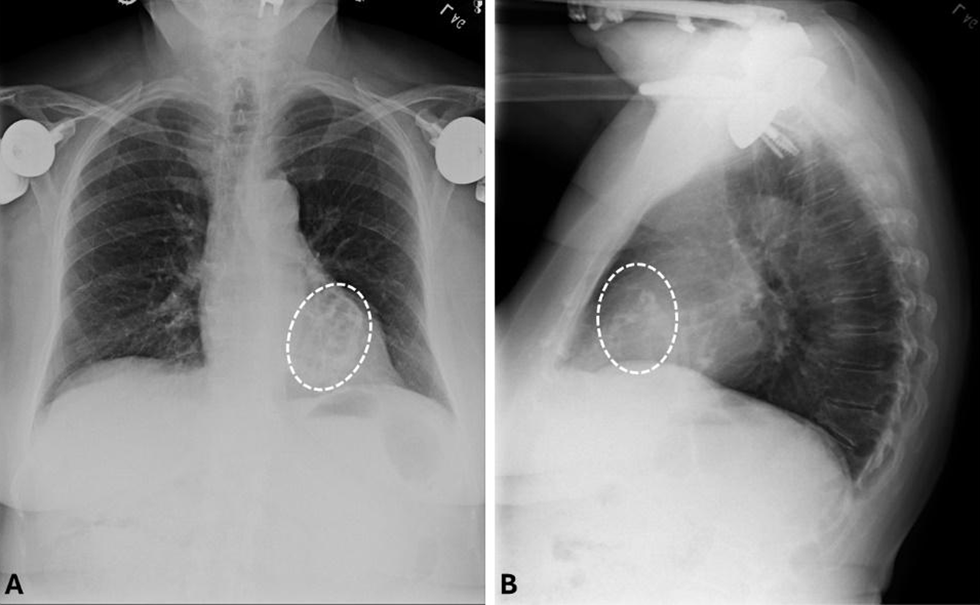
Figure 1
(A) Postero-anterior and (B) lateral radiographs clearly delineate the calcification within the heart tissue (white, dotted ovals), with the absence of hilar lymphadenopathies or other features suggestive of sarcoidosis.
Subsequently, cardiovascular magnetic resonance (CMR) was ordered and performed, which confirmed LVH and diffuse, patchy endomyocardial calcifications with peripheral hyperenhancement suggestive of myocardial scarring (Figure 2).
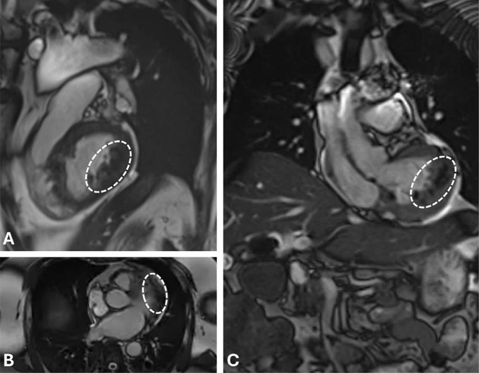
Figure 2
(A) Sagittal, (B) axial, and (C) coronal cardiac magnetic resonance imaging displays concordant hyperintensities (white, dotted ovals) supporting the diagnosis of calcification. Increased wall thickness observed clearly in panels A and C reveal the electrocardiogram-proven mild left ventricular hypertrophy.
Computed tomography angiography (CTA) with Ca scoring demonstrated no significant coronary artery stenosis (Ca Score 60) but revealed moderately extensive endomyocardial calcifications described as “metastatic” in appearance (Figure 3; Video 1).
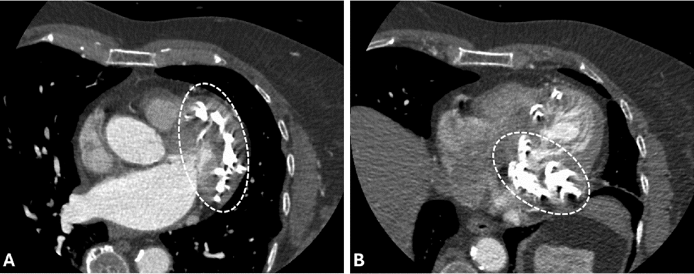
Figure 3
Axial computed tomography angiography images at the level of the (A) ascending aorta and (B) more apical aspect with hyperintensities (white, dotted ovals) spotted through the endocardial-myocardial boundaries in a diffuse pattern from apex to base.
Video 1
The axial computed tomography angiographic (CTA) examination reveals diffusely distributed hyperattenuating foci localized to the endocardial-myocardial interface, with a longitudinal distribution pattern extending from the cardiac apex to its base; see also at https://youtu.be/20ec6nuxtIA.
This comprehensive evaluation led to the diagnosis of ICC, characterized by diffuse endomyocardial calcifications in the absence of an identifiable systemic disorder. Given the patient’s stable clinical status, a conservative management approach was adopted, including continuation of antihypertensive and lipid-lowering medications, regular monitoring of heart failure progression via serial echocardiography, and patient education on medication adherence and lifestyle modifications.
Discussion
Cardiac calcification, while not uncommon, predominantly affects the pericardium rather than the other two cardiac layers. Endocardial and myocardial involvement is observed less frequently and is typically associated with metastatic deposition, infarction, or endocrine disorders.4 The etiology of endocardial calcification can be categorized into two distinct types: dystrophic and metastatic.5 Metastatic calcification is characterized by the deposition of Ca salts in previously unaffected tissues throughout the body, resulting from disruptions in calcium/phosphorus homeostasis. This phenomenon is most commonly observed in conditions such as primary hyperparathyroidism, chronic renal insufficiency, hypervitaminosis D, extensive bone destruction due to metastatic disease, or multiple myeloma.4 Conversely, dystrophic calcification represents the sequelae of localized tissue injury and cellular necrosis. While not directly associated with alterations in serum Ca levels or Ca homeostasis, the presence of hypercalcemia can exacerbate this process.6
Endomyocardial fibrosis, a rare condition of often uncertain etiology, was initially described in Brazil in 1954.7 In 1984, Silver et al. reported the first case of extensive endocardial calcification affecting the left ventricle, proposing it as a distinct entity leading to restrictive cardiomyopathy.1 This hypothesis was subsequently challenged by Lengyel et al., who emphasized that endocardial calcification was in fact indicative of endomyocardial fibrosis.8 The predominant etiology of endomyocardial calcification is frequently attributed to metastatic calcification resulting from secondary alterations in calcium and phosphorus metabolism.4 The absence of abnormalities in serum calcium and phosphorus levels excludes metastatic calcification and suggests a dystrophic process. The majority of diagnosed cases of dystrophic endomyocardial calcification and fibrosis globally are concentrated in tropical and subtropical regions, likely due to the high prevalence of tropical diseases in these areas.1 Acute cardiomyocyte injury secondary to myocardial ischemia represents another risk factor for myocardial fibrosis, which may subsequently progress to myocardial calcification.9,10 Additionally, sarcoidosis and related autoimmune disorders are recognized as potential contributory factors in the development of myocardial calcification.11
This case presents a unique diagnostic challenge due to a constellation of symptoms that align well with CAD. Cardiovascular risk factors coupled with the patient’s presentation would expect one to find clear clues on imaging such as coronary plaque calcification on CTA, wall motion defects in TTE and impaired myocardial perfusion on nuclear stress tests. While the symptom complex for CAD (angina pectoris, dyspnea on exertion) was present, no significant changes corresponded to ischemic heart disease. The ECG finding of poor R-wave progression in anterior leads, while nonspecific, was crucial in prompting further cardiac imaging to evaluate for structural abnormalities and ischemia. The detection of reversible perfusion abnormalities indicative of ischemia led to further investigation of the coronary anatomy. The TTE revealed preserved systolic function but demonstrated LVH and diastolic dysfunction—not typically associated with classic CAD.
The absence of significant coronary artery stenosis on CTA was unexpected given the ischemic changes on PET scan with discovery of endomyocardial calcifications categorized as “metastatic”—a pivotal finding that shifted the diagnostic focus. Consequently, CMR confirmed the above with peripheral hyperenhancement suggestive of myocardial scarring. This imaging modality was crucial in characterizing the extent and nature of the myocardial abnormalities. Additionally, normal serum Ca and PTH levels helped exclude common systemic causes of tissue calcification such as hyperparathyroidism or renal pathologies. The absence of significant CAD, normal calcium metabolism, and lack of evidence for systemic inflammatory disorders led to the diagnosis of ICC.
Conclusion
This case demonstrates an intriguing instance of incidentally discovered endomyocardial calcification. The presentation challenges the typical understanding of endomyocardial fibrosis and calcification, which are often associated with symptomatic heart failure and more commonly found in tropical and subtropical regions. Despite findings indicating the presence of CAD, the disproportionate amount and location of calcification and fibrosis found in other imaging modalities was unequivocal in ruling out CAD as the cause.
By virtue of the report presented, we also underscore the importance of a systematic and comprehensive diagnostic approach in patients presenting with atypical features of common cardiac conditions. This case highlights the value of multimodality cardiac imaging in uncovering rare pathologies and emphasizes the need for individualized management strategies in complex cardiac cases. Further research is needed to better understand the etiology, natural history, and optimal management of ICC.
Key Points
This case highlights the importance of multimodality imaging, which is vital in detecting subtle or asymptomatic cardiac abnormalities.
Both metastatic and dystrophic causes of endomyocardial calcification must be considered, even in patients from non-tropical regions.
Competing interests
The authors have no competing interests to declare.