
Case Description
An 80-year-old female presented to the emergency department with worsening shortness of breath. She had a complex medical history including severe aortic stenosis (AS) status post transcatheter aortic valve replacement (TAVR) in 2017, followed by a valve-in-valve (ViV) in 2021 due to valve degeneration, chronic obstructive pulmonary disease with bronchiectasis (on chronic 2-4 L oxygen), coronary artery disease with percutaneous coronary intervention to the left anterior descending artery, chronic diastolic heart failure, and atrial fibrillation on apixaban. She reported intermittent non-exertional chest pain exacerbated by breathing but denied other associated symptoms. She was desaturating to 82% on arrival, which improved with 6 L non-rebreather mask. A transthoracic echocardiogram revealed severe stenosis of the prosthetic aortic valve with a mean gradient of 49 mm Hg and an orifice area of 0.75 cm². Given the severity of her symptoms and the severity of the valve obstruction, cardiothoracic surgery was consulted and planned for surgical correction. Following comprehensive evaluation of the case, the decision was made to perform an explantation of her two existing valves (an initial, outer 26-mm CoreValve Evolut R TAV® [Medtronic] and an inner 23-mm Sapien 3 ViV® [Edwards Lifesciences]) with removal of the CoreValve leaflets and deployment of a new TAV within the CoreValve stent frame under direct vision.
Following sternotomy and cardiopulmonary bypass (CPB) initiation, the ascending aorta was opened transversely above the CoreValve valve stent frame. The Sapien valve was examined and appeared severely stenotic with heavily calcified leaflets. The stent frame of the Sapien was carefully crushed and removed, allowing full exposure of the underlying CoreValve valve. The CoreValve was also severely calcified and obstructed, especially at the right and left coronary ostia. The CoreValve leaflets were resected in multiple pieces to fully visualize the coronary ostia (Figure 1).
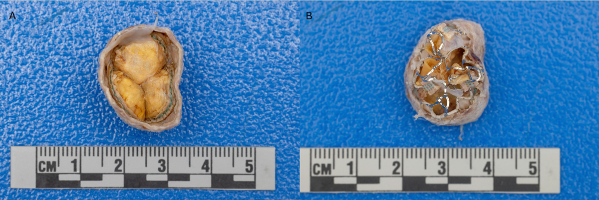
Figure 1
Explanted Sapien aortic valve with (A) superior view, (B) inferior view.
A new 23-mm Sapien Ultra Resilia® (Edwards Lifesciences) valve was then prepared and positioned inside the stent frame of the CoreValve. The new valve was deployed and slowly inflated under direct vision with the Edwards Certitude Delivery System® (Edwards Lifesciences), with the distal portion of the valve stent below the left coronary ostium to avoid interference with the ostia. The aortic cross-clamp was removed after 47 minutes, and the patient was weaned off CPB after 73 minutes. Post-CPB transesophageal echocardiogram showed no evidence of aortic insufficiency, with trivial periprosthetic regurgitation and a gradient of 5 mm Hg. Hemostasis was achieved, the sternum was closed, and the patient was transferred to the intensive care unit in stable condition.
The patient’s recovery was uneventful. On postoperative day (POD) 1, the patient was extubated, and vasopressors and dobutamine were weaned off on POD 3 and 7, respectively. By POD 10, she was deemed stable for discharge to a skilled nursing facility on 4 L of home oxygen with warfarin for anticoagulation. At 1-month follow-up, the patient was doing well on her baseline oxygen requirements without shortness of breath and with a well-healing sternal incision.
Discussion
TAVRs have become very popular and widely utilized over the past decade.1 Bioprosthetic valves can calcify and degrade over time, requiring replacement around 10 to 15 years after initial placement depending on the patient’s risk factors.2 Occasionally, premature valve dysfunction due to improper placement or sizing issues can require earlier valve replacement.3 There are two main approaches to TAVR reintervention: (1) redo-TAVR, or “TAVR-in-TAVR”: transcatheter placement of a new aortic valve within the previous, dysfunctional valve; and (2) TAVR-explant, or “SAVR-after-TAVR”: surgical removal of the dysfunctional transcatheter aortic followed by surgical aortic valve replacement (SAVR).
Complicating factors, such as endocarditis or a history of TAVR-in-TAVR, can affect the decision to pursue surgical explantation over valve-in-valve replacement. The most frequent indications for TAVR-explant are structural valve degeneration (SVD), aortic insufficiency/paravalvular leaks, and index TAVR procedural complications.4 Analysis of databases from the Society of Thoracic Surgeons (STS) and the American College of Cardiology estimate the incidence of TAVR-explant to be 0.4% to 1% of index (initial) TAVR placements.1,3 This is, however, the fastest growing cardiac surgical procedure in the US.5
While TAVR has gained popularity and reputation as a safe and effective procedure for a wide patient population, TAVR failures can carry significant risks. Over two-thirds of TAVR-explant procedures require concomitant cardiac surgical procedures, most of which involve aortic root reconstruction from aortic damage during the explantation.1,3,4 As a result of these extra surgical risks, the operative mortality of TAVR explantation triples compared to index procedures1 and rises from 17.1% to 41.2% when concomitant aortic repair is performed.3,4 However, it is also important to acknowledge the lack of individual surgeon experience in this recently growing procedure and the limited data on optimal approach.
Per the 2020 STS database analysis, the median number of explants performed per surgeon and center was one for both.3 In light of our experience as a quaternary referral center, these numbers have grown and have likely contributed to the drop in operative mortality rates over the years from 25% to 30% to less than 15% in more recent studies.6,7 In addition to concomitant aortic root repair, other frequently performed procedures during the explant, such as coronary artery bypass grafting (13.8-17.7%) or mitral valve repair (21.6-23.2%), can also bring additional risks to the explant and, in certain studies, have been reported to be more commonly performed than the root replacements/repair.6,8 The learning curve in addition to the patient’s higher risk profile likely contributes to the complication rates of TAVR explants rather than the procedure itself.
In this elderly, high-operative-risk patient with multiple comorbidities, the decision to preserve the inner valve stent frame and deploy a TAVR directly into the field minimized interactions with the aortic wall, which also reduced the risk of damage that could have ensued from complete bioprosthesis explantation and a consequent SAVR. Concomitant aortic root reconstruction can occur in 13.4% to 18.8% of TAVR explants per the literature.6,8 This is likely due to the endothelization of the aorta into the stent-frame of the valve that makes the TAVR explant challenging, where layers of aortic tissue must be carefully dissected from the nitinol cage without damaging the aortic wall. In our case, the aortic cavity was already exposed, and the new valve was easily aligned and deployed under direct visualization. The new valve was positioned below the coronary ostia to facilitate adequate coronary flow, which was initially obstructed with the old bioprosthesis, as well as provide access should another TAV be needed in the future.
The Resilia valve was chosen due to its favorable profile of 99.3% freedom from SVD and 97.2% freedom from reoperation per the COMMENCE aortic trial, although its long-term durability is still to be explored as the mean follow-up time was 7.7 years.9 Nonetheless, routine monitoring in this patient is warranted, given her history of accelerated bioprosthetic valve degeneration. Positioning the new valve so that a future TAVR could be performed, if necessary, helped balance the need to minimize operative risk through this technique with the concerns for valve durability.
Pirelli et al. performed a similar approach in 2021 to surgically explant a TAVR and perform a TAVR under direct vision through the aortotomy, which was termed Surgical Resection of Prosthetic Valve Leaflets Under Direct Vision (SURPLUS).10 We present the first known case of a hybrid surgical-interventional technique being used in a patient with multiple prosthetic TAVR explantations with an excellent intra- and postoperative course. Kelly et al. and Liang et al. present case reports of multiple TAVR explants; however, all TAVRs were completely explanted, and SAVR was performed in both cases with concomitant aortic repair.11,12
The presented technique, a partial TAV explantation with a consequent TAVR done under direct visualization, has several advantages to traditional TAVR explant approaches, including more precise valve positioning with the direct vision, less time spent on cardiopulmonary bypass, and lower risk of aortic damage, which can be a large driver of mortality in these cases. However, the approach is not without limitation. SURPLUS may not be suitable in cases where explantation of the entire valve is necessary, such as when infective endocarditis is present. In addition, while the approach may be favorable for elderly or high-risk patients, it may not be a good option for younger, more active patients. Younger TAVR patients generally exhibit limited bioprosthetic valve durability due to their longer lifespans, with higher rates of SVD and patient-prosthesis mismatch (PPM) in the long run.13 If PPM was present, it would not be correctable using the SURPLUS technique, since the retained stent frame approach prevents annular enlargement.
Conclusion
Using catheter technology under direct vision for complex TAVR re-interventions could potentially decrease the risk for concomitant cardiac procedures, which might contribute to lower operative mortality. As TAVR procedures expand to younger populations and surgeons gain more experience with TAVR re-intervention, new technologies and innovative surgical approaches will continue to improve TAVR safety and complication management with the hope to optimize long-term outcomes.
Competing Interests
The authors have no competing interests to declare.