
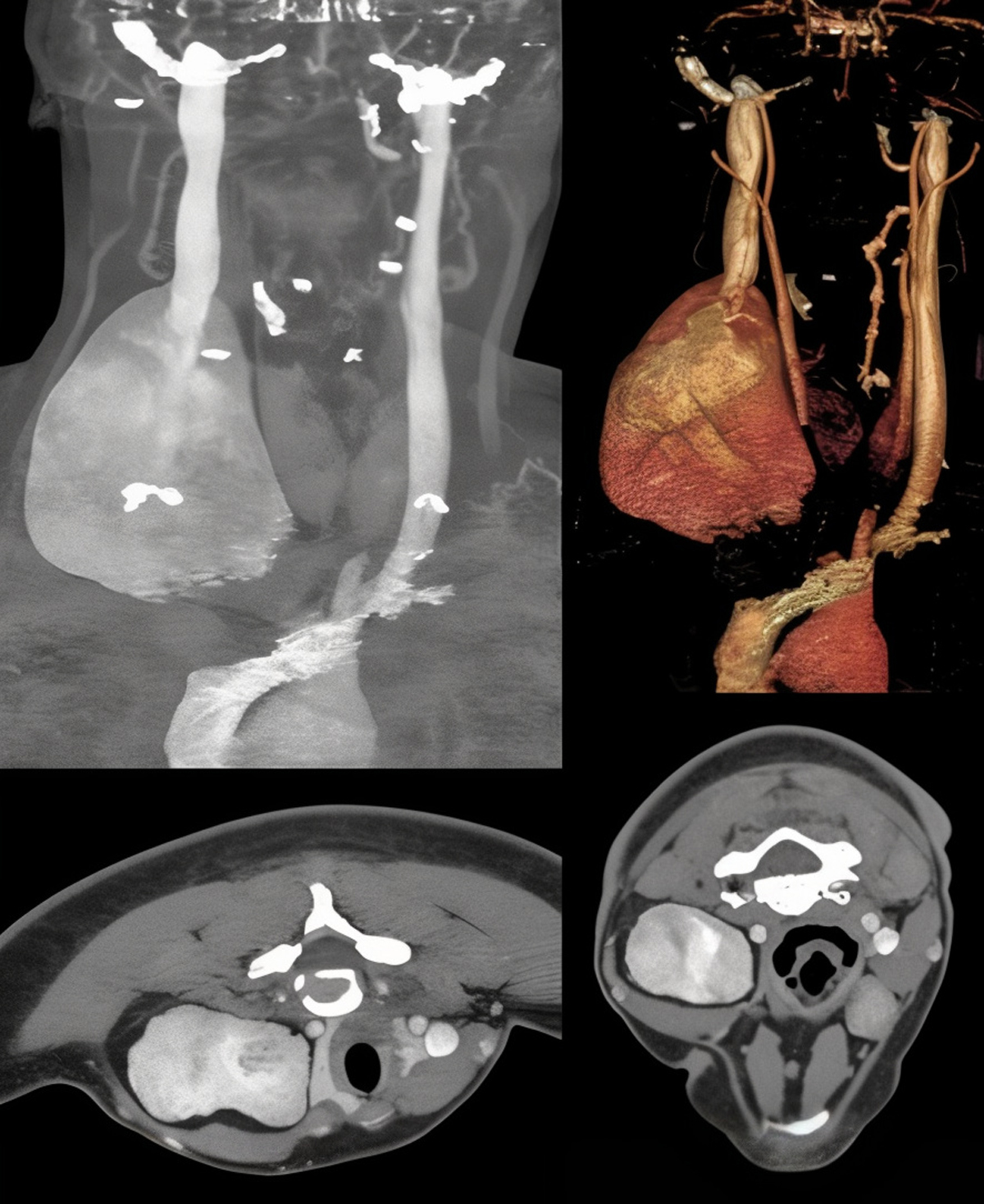
Figure 1
Preoperative computed tomography cerebral venography imaging of the internal jugular vein aneurysm.
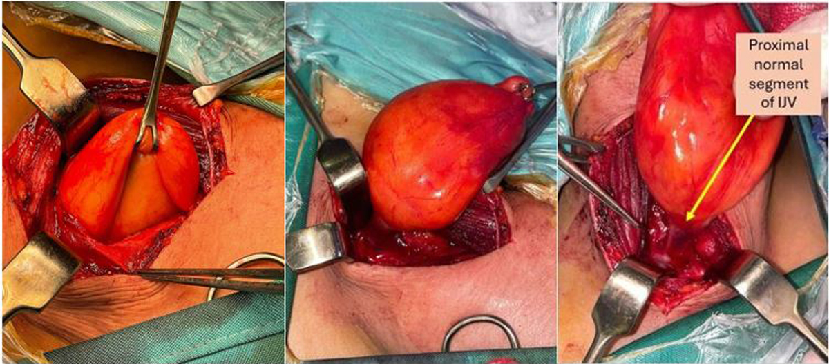
Figure 2
Intraoperative presentation of the internal jugular vein (IJV) aneurysm, and excising the vein up to the proximal segment of IJV. Arrow denotes the proximal normal segment of the IJV.
Video 1
Demonstration of transmitted respiratory flow within the aneurysmal segment of the internal jugular vein. The video highlights the movement of the venous wall synchronized with the patient’s breathing pattern; see also at https://youtube.com/shorts/gbH9sNRZDcM.
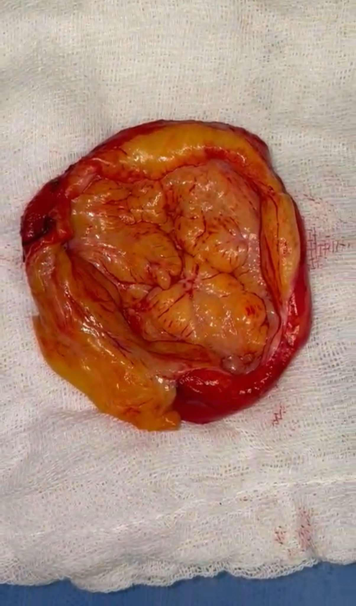
Figure 3
Content of the aneurysm.
Video 2
Surgical opening of the aneurysmal segment. The video captures the intraoperative exploration of the aneurysm’s content, revealing the fibrolipomatous structure and confirming the absence of luminal occlusion; see also at https://youtube.com/shorts/tgAK2F-QTCY.