
Video 1
Intraoperative recording of the surgery, fluoroscopy, and transcranial Doppler monitoring; see also at https://www.youtube.com/watch?v=cRSKdlaTNSQ&t=93s.
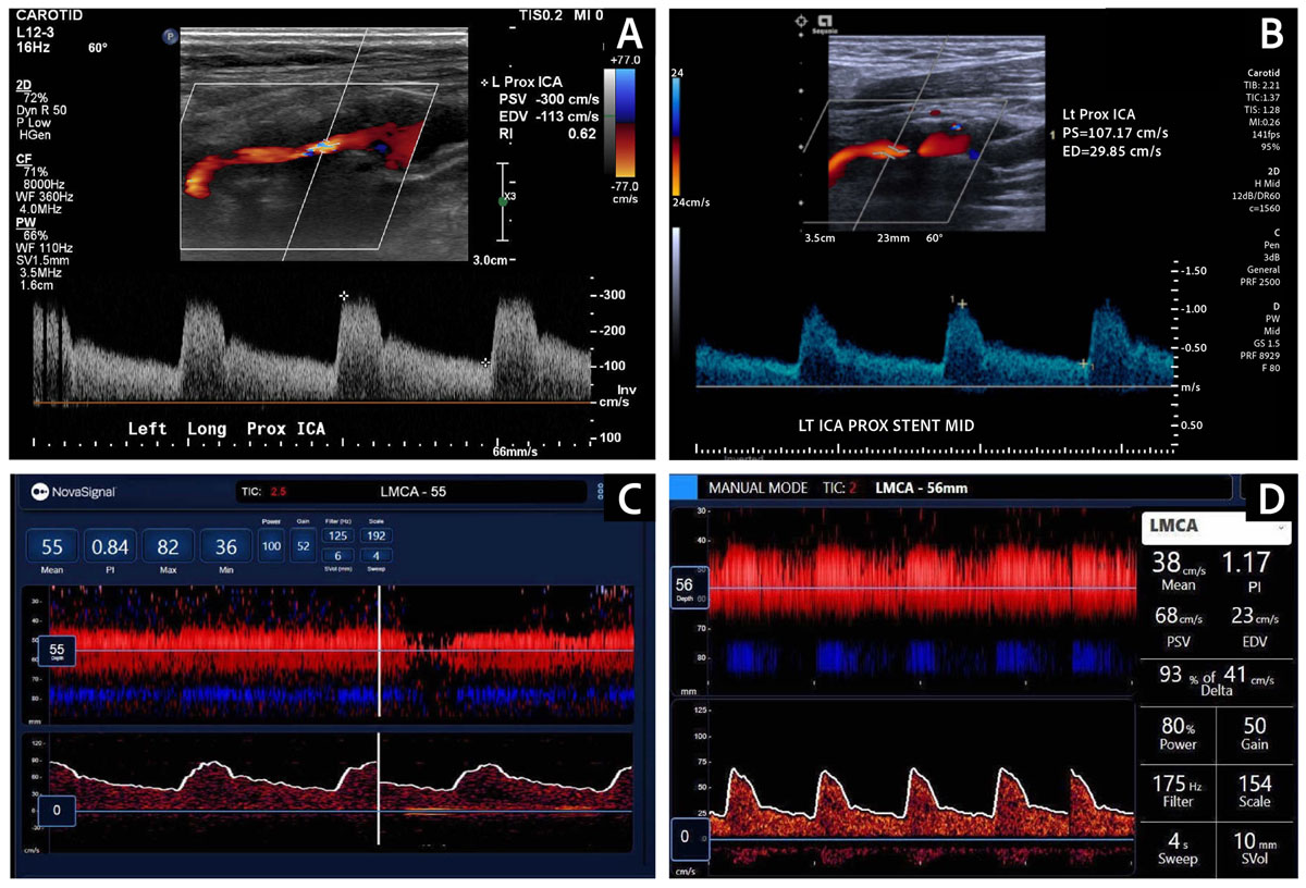
Figure 1
(A) Preoperative carotid Duplex ultrasound revealed severe stenosis in the proximal ICA (PSV of 300 cm/sec, EDV of 113 cm/sec). (B) Postoperative carotid Duplex ultrasound confirmed good stent patency, normal flow velocities (PSV of 107.17 cm/sec, EDV of 29.85 cm/sec) and improved pulse waveforms. (C) Preoperative TCD showed a blunted left MCA pulse waveform, confirming a proximal stenosis. (D) TCD after stenting revealed substantial improvement in the systolic upstroke in the pulse waveform of the left MCA. ICA: internal carotid artery; PSV: peak systolic velocity; EDV: end diastolic velocity; TCD: transcranial Doppler; MCA: middle cerebral artery
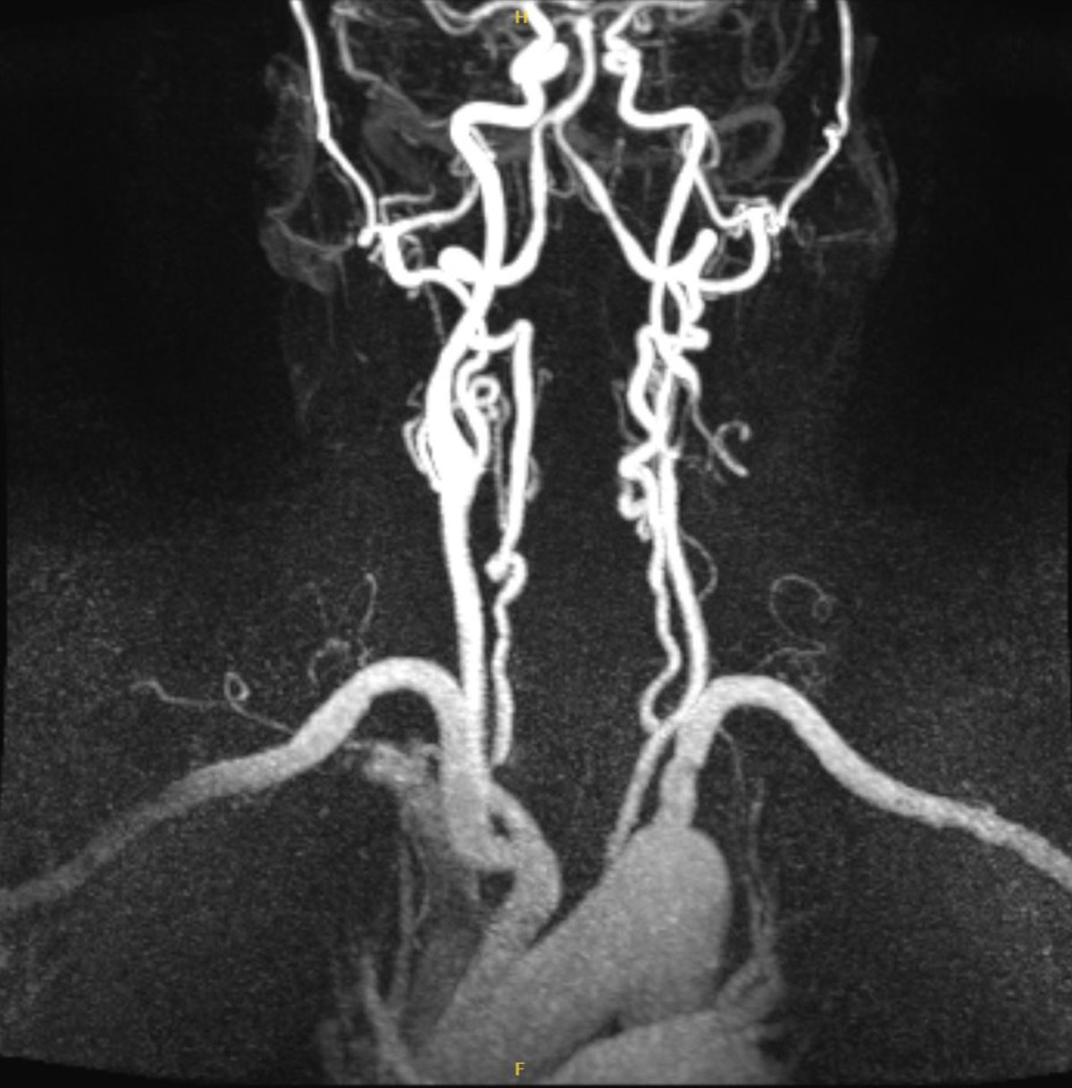
Figure 2
Magnetic resonance angiogram revealed significant left internal carotid artery stenosis (70-80%).
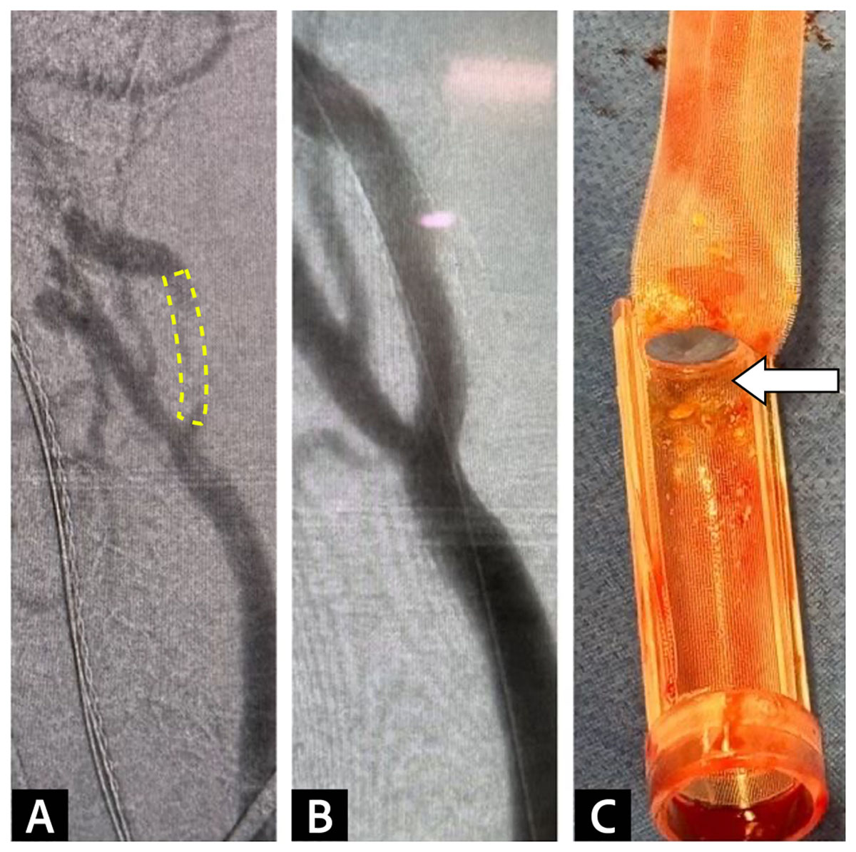
Figure 3
(A) Angiogram before stenting demonstrated stenosis in the internal carotid artery. (B) Post-stenting angiogram confirmed stenosis resolution. (C) The 200-micron filter integrated into the flow reversal system captured embolic debris before it reached the venous return sheet.