
Case Presentation
A 26-year-old male patient with a smoking history of 12 pack years presented to the emergency department with atypical chest pain. He had exertional chest pain and dyspnea for the past 2 years. No effort-related syncope or palpitations were reported, no significant medical issues or regular medication use were reported, and there was no family history of cardiovascular disease. He works as a merchandiser and engages in regular physical activities.
Physical examination, including his vital signs, were unremarkable. The initial evaluation aimed to rule out acute coronary syndrome by performing an electrocardiogram and serial cardiac troponin levels, which were negative. Following this, 2-dimensional transthoracic echocardiography was performed, showing normal structure and function of all four cardiac chambers and valves.
Subsequent computed tomography angiography revealed a complete absence of the right coronary artery (RCA) at the level of the right coronary ostium (Figure 1). The heart exhibits a single coronary artery (SCA) as the RCA arises from the distal left circumflex artery (LCx) (Figures 2, 3; Video 1). No evidence of stenosis or atherosclerosis was observed in any of the coronary arteries.
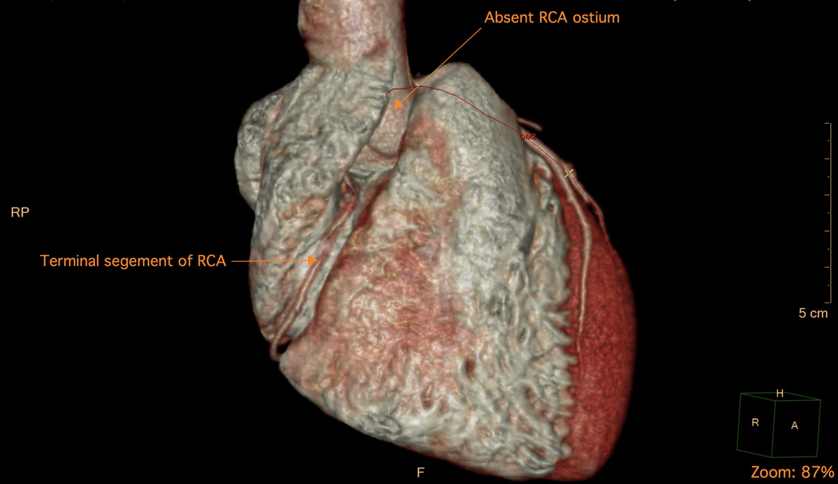
Figure 1
Cardiac computed tomography angiography 3-dimensional reconstruction showing absent right coronary artery (RCA) ostium and terminal segment of RCA.
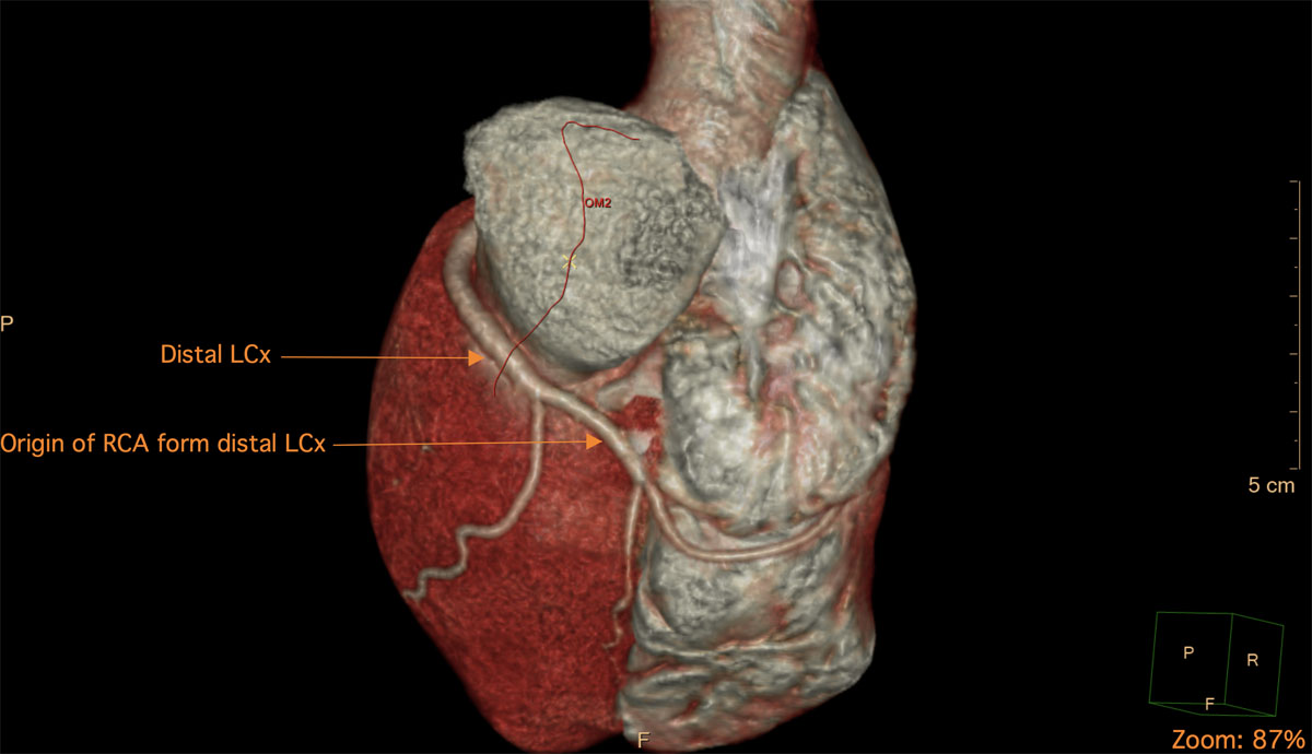
Figure 2
Cardiac computed tomography angiography 3-dimensional reconstruction showing right coronary artery originating from the left circumflex artery. RCA: right coronary artery; LCx: left circumflex artery
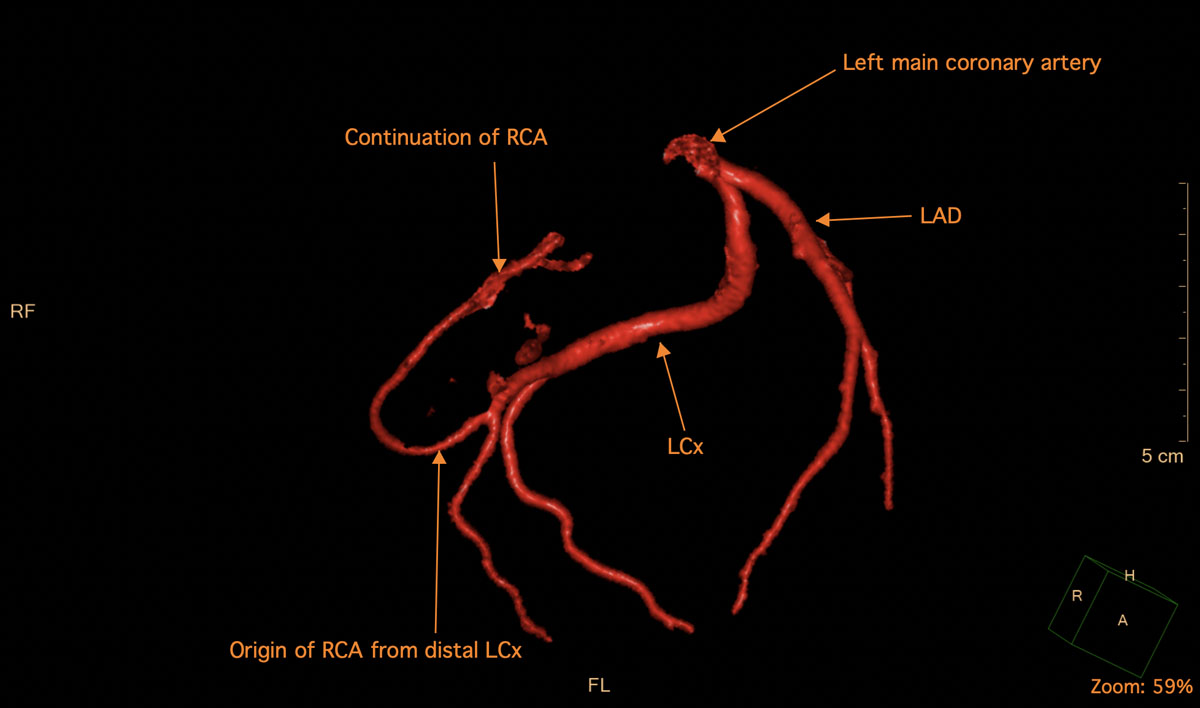
Figure 3
Coronary computed tomography angiography-subtracted VRT reconstruction image shows origin of right coronary artery (RCA) from left circumflex artery with absence of RCA ostium. VRT: volume rendering technique; LCx: left circumflex artery; LAD: left anterior descending artery
Video 1
Cardiac computed tomography angiography 3-dimensional reconstruction shows the complete absence of right coronary artery (RCA) ostium and the course of RCA as it arises from the left circumflex artery (LCx) and terminates in the right coronary sulcus; see also at https://youtu.be/gfDtCQCge00.
We offered the patient cardiac nuclear testing to evaluate microvascular ischemia and myocardial blood flow reserve. However, he was reluctant due to concern about radiation exposure. Our management strategy was conservative, emphasizing strict lifestyle modifications including smoking cessation, regular lipid profile evaluations, and the avoidance of strenuous exercise.
Discussion
According to the classification proposed by Lipton-Yamanaka et al., the described case is an LI type: (L) indicates that an SCA arises from the left cusp, giving off the left anterior descending and LCx, with the RCA originating from the LCx; (I) indicates that all the mentioned arteries follow their normal courses.1 Patients with SCA can present with cardiomyopathy or acute coronary syndromes, but this particular anomaly was reported to have a benign course, with negative cardiac work-up in the majority of cases unless there is significant atherosclerosis.2 Some case reports attributed exertional chest pain to abnormally slow blood flow to the RCA. This reasoning was supported by the patient’s excellent response to medical management with calcium channel blockers and nitrates and the absence of significant atherosclerosis on work-up.3 True right myocardial ischemia that required primary coronary intervention was reported in a case as well,4 while another case was incidentally found while working up pulmonary embolism.5 In conclusion, the presentation for this anomaly is highly variable, and treatment strategy is tailored to a case-by-case basis. Percutaneous coronary intervention providers should carefully review primary team imaging to facilitate easier manipulation and reduce contrast usage, thereby enhancing patient care.
Competing Interests
The authors have no competing interests to declare. The patient provided informed consent for documenting this case.