
Video 1
Transesophageal echocardiography showing a thrombus in the left upper pulmonary vein in the presence of MitraClip; see also at https://youtu.be/ukYRitIx3_8.
Video 2
Transesophageal echocardiography with color Doppler showing a thrombus in the left upper pulmonary vein in the presence of MitraClip; see also at https://youtu.be/NsPPSGp1BiI.
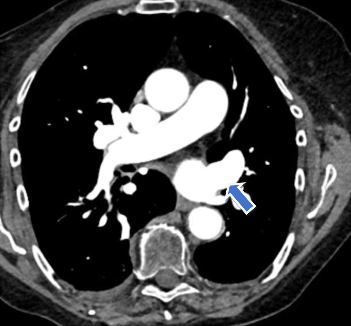
Figure 1
Computed tomography of the chest showing no thrombus in the left upper pulmonary vein.
Table 1
Case timeline. TEER: transcatheter edge-to-edge repair; X: day of mitral valve TEER; TEE: transesophageal echocardiogram; MR: mitral regurgitation; TR: tricuspid regurgitation; CT: computed tomography
| CASE TIMELINE | |
|---|---|
| X – 48 months | Underwent pulmonary vein isolation ablation for atrial fibrillation |
| X – 2 months | TEE before mitral valve TEER showed severe MR (2–3+), severe TR, and no thrombus in the left atrium appendage or pulmonary veins |
| X – 3 days | Rivaroxaban was paused for 3 days before mitral valve TEER |
| X | Underwent mitral valve TEER for severe MR. TTE after mitral valve TEER showed a reduction of MR (2–3+ to 1+), mean mitral valve gradient 2 mm Hg, no thrombus in the left atrium appendage or pulmonary veins. Rivaroxaban resumed the next day |
| X + 2 months | Day of Watchman placement: TEE showed a thrombus in the left superior pulmonary vein. Watchman placement was aborted, and the patient was switched from rivaroxaban to apixaban |
| X + 3.5 months | CT of the chest showed resolution of the thrombus in the left superior pulmonary vein while on apixaban for 6 weeks |