
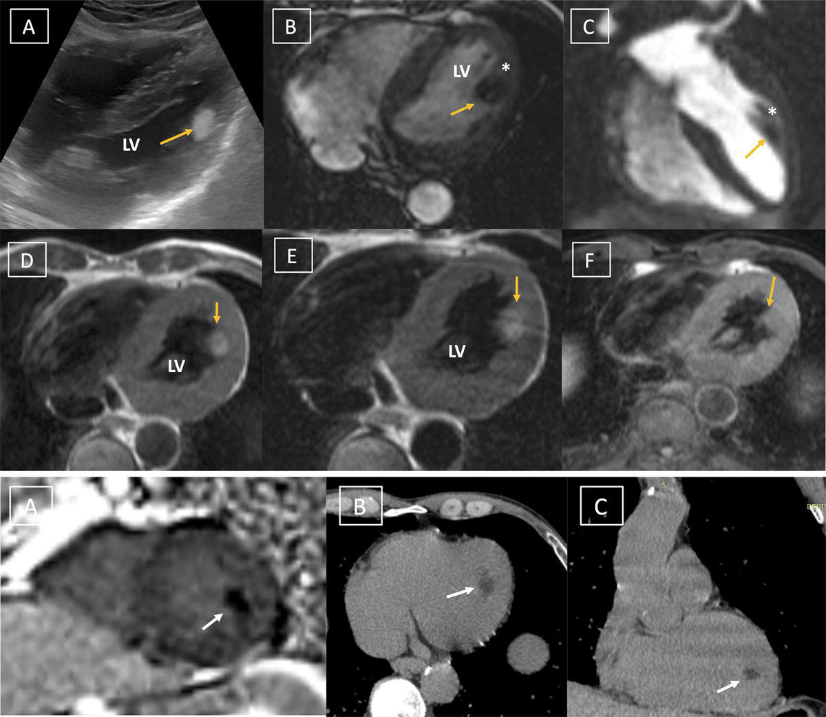
Figure 1
(A) Transthoracic echocardiography reveals echogenic oval mass (depicted with yellow arrow) with regular margins attached to the lateral myocardial wall of the left ventricle. (B) Cine steady state free precession four-chamber image demonstrating oval mass attached to the papillary muscle (white asterisk). (C) Four-chamber image clearly depicting the hypointense mass attached to anterolateral papillary muscle (white asterisk). The mass is hyperintense on (D) T1 and (E) T2 weighted image with suppression on (F) fat saturated sequence suggesting a cardiac lipoma.
Figure 2 (A) Short axis late gadolinium enhanced image demonstrating no enhancement within the mass. (B) Axial and (C) coronal computed tomography image depicts a hypointense mass (white arrow) with fat attenuation suggesting cardiac lipoma.