
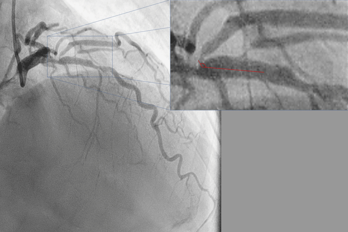
Figure 1
Diagnostic angiogram in right anterior oblique 40 cranial 40 view using a 6F catheter (radial access) showing a true bifurcation stenosis of the left anterior descending (LAD) diagonal junction. The ostium of the large diagonal is diseased and arises from the LAD at an angle of approximately 130° with the main branch (inset).
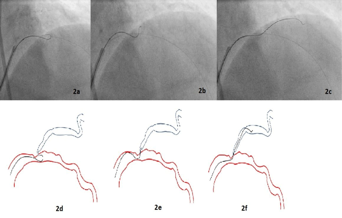
Figure 2
Angiogram images (2a-c) of percutaneous intervention (2a) through the femoral route showing advancement of the wire beyond the tip of the micro-catheter such that the wire assumed its pre-formed shape; (2b) the micro-catheter was withdrawn back into the guiding catheter, and with careful rotational manipulation the wire was pulled back until its tip engaged the ostium of the diagonal branch; (2c) once the wire tip entered the diagonal, the wire was advanced while torquing. Line diagram (2d-f) depicting the same information.
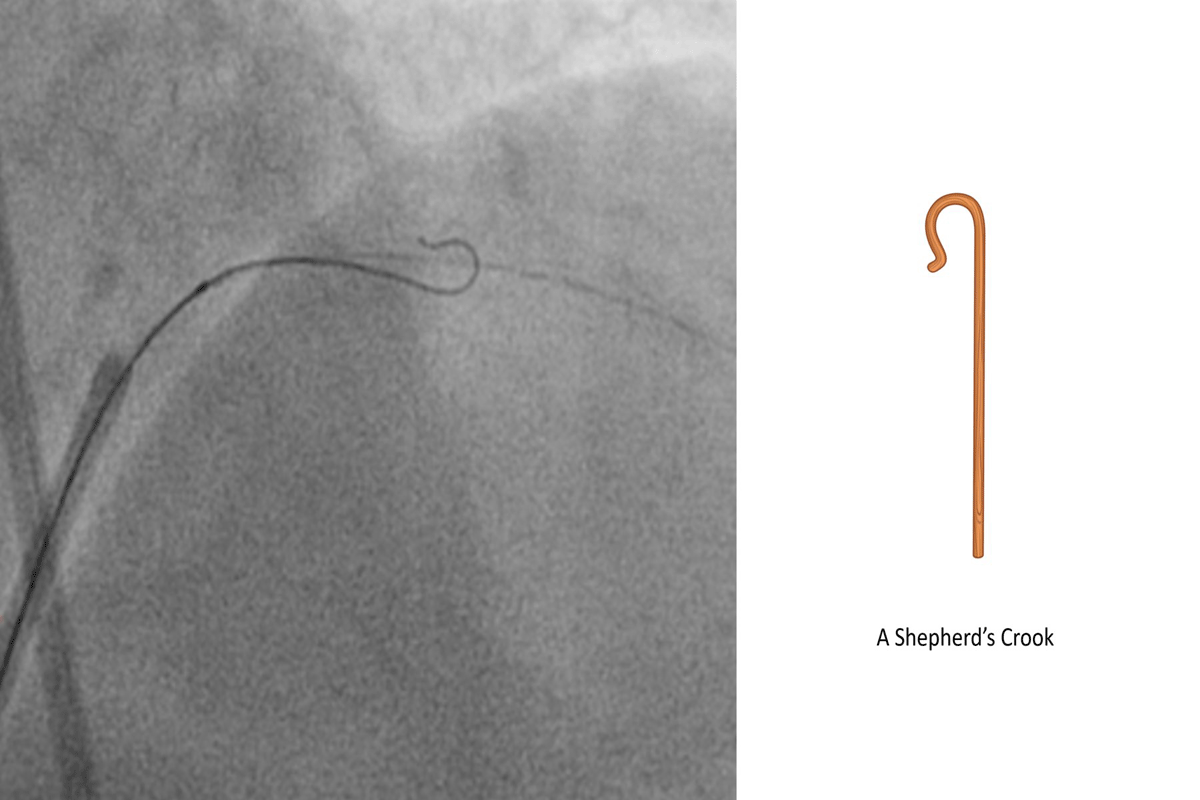
Figure 3
Diagnostic angiogram and line diagram image showing shepherd’s crook curve: beginning approximately 6 mm from the tip, a bend of 180° opposite the direction of the primary curve was introduced into the wire.
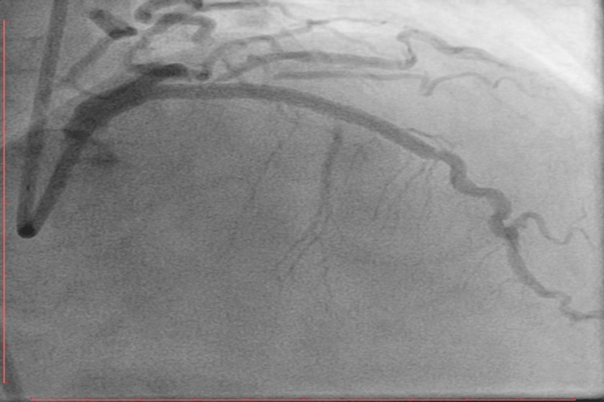
Figure 4
An angiogram in the right anterior oblique 40 cranial 40 view shows the final TIMI III flow in the left anterior descending artery after stenting and post dilatation with a non-compliant balloon.