
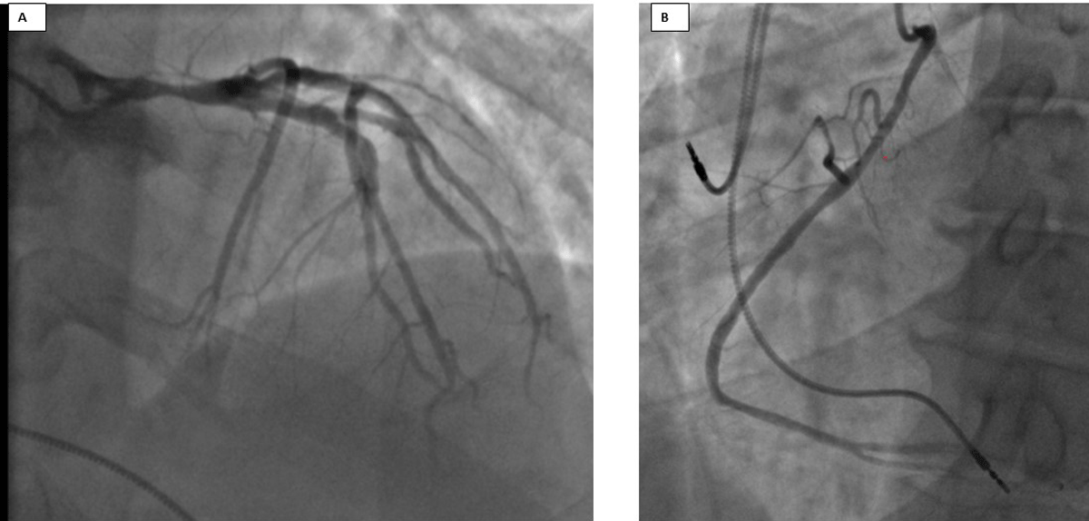
Figure 1
Initial coronary angiogram. (A) 80% stenosis of the left main artery, 40% stenosis in the proximal left anterior descending artery. (B) Right coronary artery had an anomalous origin from the ascending aorta.
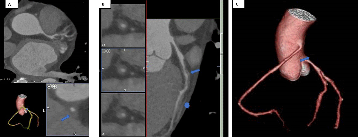
Figure 2
Coronary computed tomographic angiography (CCTA). (A) Diffuse narrowing of the left main artery with 70-80% maximal stenosis; CCTA reformat depicts diffuse narrowing of the left main artery resulting in 40-50% stenosis (arrow). (B) Curve planar reformat of the left anterior descending (LAD) shows diffuse circumferential wall thickening of the proximal LAD resulting in up to 50% stenosis (arrow). Incidental 1.4-cm myocardial bridge involves the mid-LAD without extrinsic compression or significant intraluminal stenosis (*). (C) 3-dimensional volume rendering of the coronary tree demonstrates high takeoff of the right coronary artery from the anterior surface of the ascending aorta (arrow).
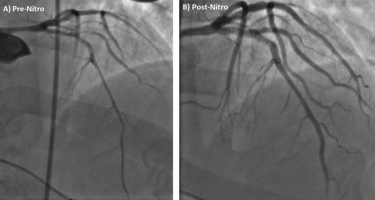
Figure 3
Repeat coronary angiogram. Pre-nitroglycerine coronary angiogram with changed severity and distribution of left main artery (LMA) stenosis compared to recent prior coronary angiogram (Figure 2). Coronary angiogram after intracoronary nitroglycerin administration with improvement in LMA stenosis and improved downstream blood flow.
Video 1
Initial coronary angiogram pre-intracoronary nitroglycerin administration; see also at https://youtu.be/3oVymC12vP8.
Video 2
Repeat coronary angiogram after intracoronary nitroglycerin; see also at https://youtu.be/pXcMcIz8Vkw.
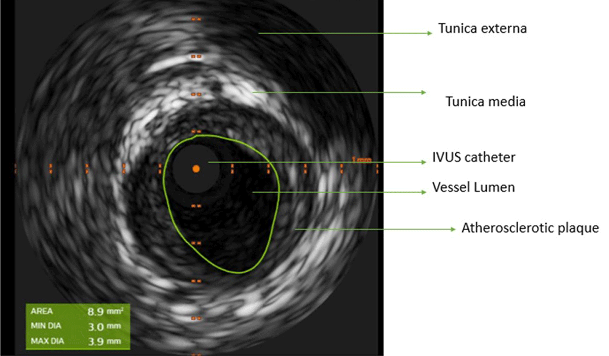
Figure 4
Intravascular ultrasound (IVUS) of the left main artery showing minimal luminal area of 8.9 mm2 and mild-moderate plaque burden.
Table 1
| SPONTANEOUS EPISODE | PROVOKED EPISODE |
|---|---|
| Nitrate-responsive angina + transient ischemic electrocardiographic (ECG) changes or angiography documented coronary artery spasm (defined as > 90% coronary artery occlusion spontaneously) | Nitrate-responsive angina + transient ischemic ECG changes + angiography documented coronary artery spasm (defined as > 90% coronary artery occlusion with provocation) |
Table 2
| Avoid precipitants | Smoking Sympathomimetics – cocaine, methamphetamine, ecstasy, adrenaline Other agents – beta-blockers, ergot alkaloids Mental stress |
| Calcium channel blockers (first-line therapy) | Non-dihydropyridine (verapamil, diltiazem), dihydropyridine (nifedipine, amlodipine) |
| Antianginal therapy | Nitrates, nicorandil, statins, cilostazol |
| Refractory angina | Stellate ganglion block |
Table 3
Prognostic factors in vasospastic angina.
| Poor prognostic factors in vasospastic angina | Serious clinical presentations (ST-elevation, acute coronary syndrome, cardiac arrest), combined atherosclerotic stenosis, multi-vessel spasm, not using calcium-channel blockers, definite spasm on provocative testing, focal spasm |