
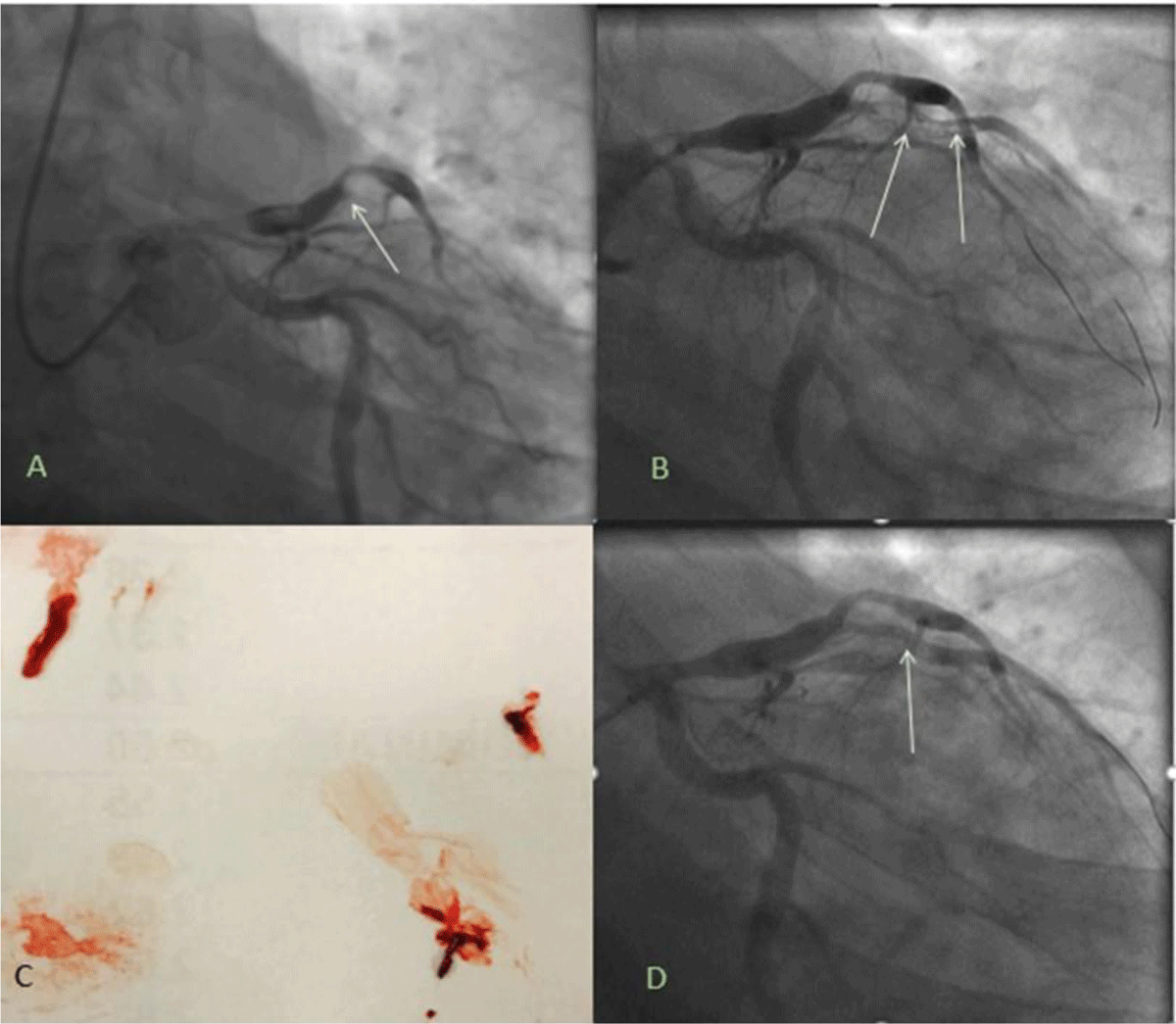
Figure 1
(A) Acute thrombotic occlusion of first diagonal (D1 vessel (white arrow). (B) Coronary guidewires advanced into distal vessels and balloon angioplasty performed delineating the vessel and revealing a large thrombus burden (white arrows). (C) Aspiration thrombectomy with large thrombus retrieval. (D) Angiography after intracoronary tissue plasminogen activator and Abciximab revealed partial thrombus resolution but still with a mobile thrombus (white arrow).
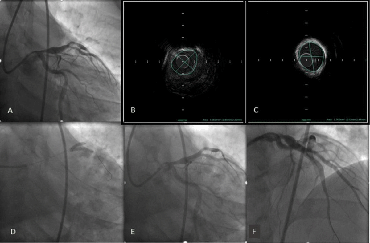
Figure 2
(A) Angiography 48 hours later showed near thrombus resolution. (B) Intravascular ultrasound of the mid-left anterior descending artery (C) and first diagonal branch (D1) showed minimal lumen area of 3.7 mm2 and 3.4 mm2, respectively. (D) Simultaneous kissing stent placement in the left anterior descending artery and the D1 branch. (E, F) Final angiography revealed thrombolysis in myocardial infarction-3 flow and no residual stenosis.
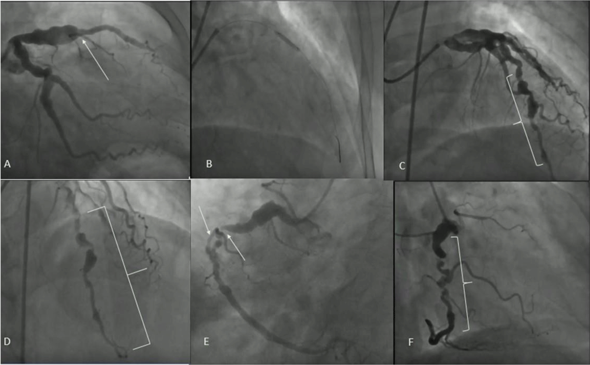
Figure 3
(A) Aneurysm of the proximal left anterior descending artery (LAD) with acute thrombotic occlusion of the mid-LAD with thrombolysis in myocardial infarction (TIMI)–0 flow distally. (B) Stent placement. (C, D) Successful revascularization with restoration of TIMI-3 flow. (E,F) Aneurysm in the proximal right coronary artery (RCA) with no significant disease. Mid-RCA had a focal 90% lesion with a tortuous course afterwards and TIMI-3 flow distally.
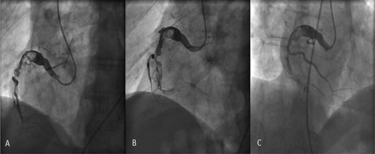
Figure 4
(A) Aneurysmal dominant right coronary artery (RCA) with a large thrombus burden and a thrombotic occlusion of the mid RCA. (B, C) Aspiration thrombectomy achieving only partial flow in the distal vessel.
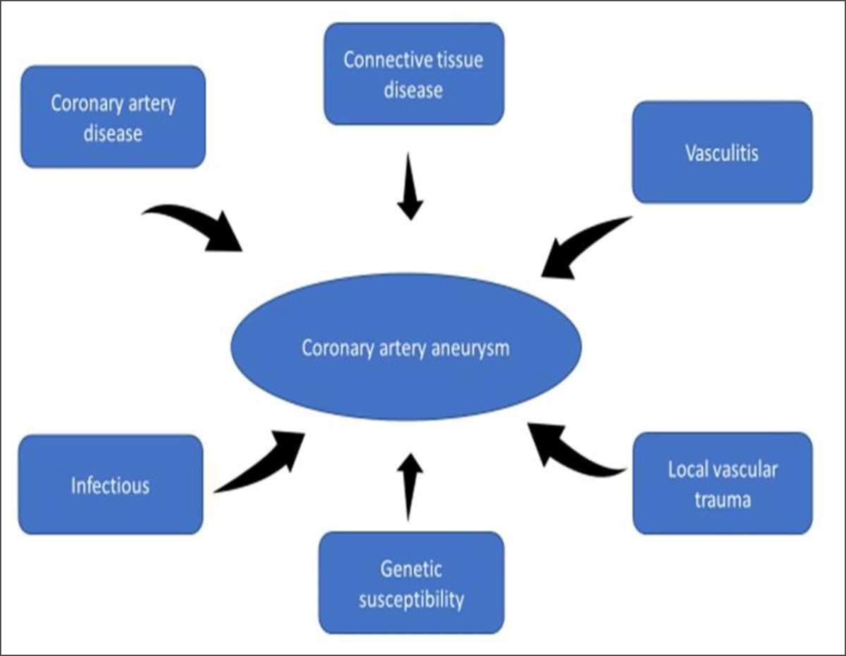
Figure 5
Etiologic mechanisms for coronary artery aneurysms.