
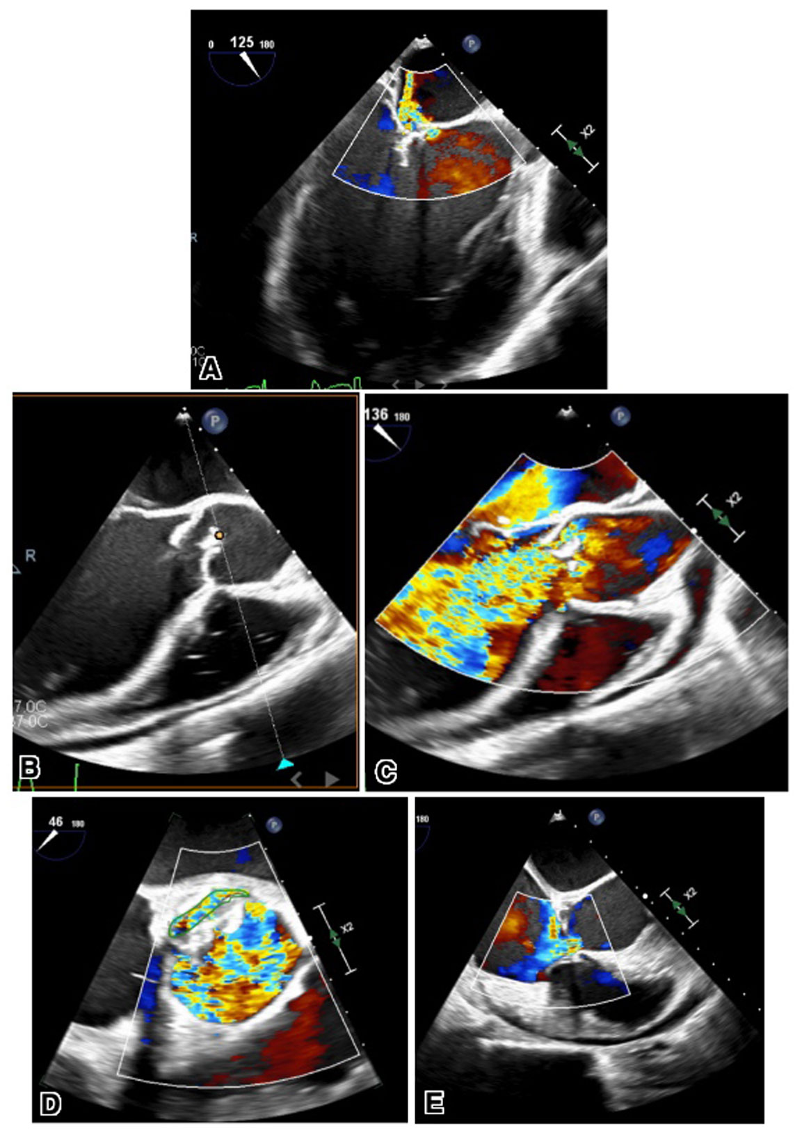
Figure 1
(A) Transesophageal echocardiogram images showing perforated anterior leaflet of the mitral valve. (B) Long- and short-axis views showing perforated aortic valve (C, D) with color flow Doppler. (E) Transesophageal echocardiography long-axis view with color flow Doppler showing ventriculoatrial shunt flow from the left ventricle into the right atrium.
Video 1
Two-dimensional transesophageal echocardiography long-axis view showing perforated anterior leaflet of the mitral valve; see also at https://youtu.be/pEjZ2OEgRFw.
Video 2
Two-dimensional transesophageal echocardiography long-axis view showing perforated aortic valve; see also at https://youtube.com/shorts/JyoJwJF3QJM.
Video 3
Two-dimensional transesophageal echocardiography long-axis view with color flow Doppler showing perforated aortic valve; see also at https://youtu.be/cYE_BPoucqE.
Video 4
Two-dimensional transesophageal echocardiography short-axis view with color flow Doppler showing perforated aortic valve; see also at https://youtu.be/HrZUch23SbA.
Video 5
Two-dimensional transesophageal echocardiography long-axis view with color flow Doppler showing ventriculoatrial shunt flow from the left ventricle into the right atrium; see also at https://youtu.be/LQRjok6eTGM.