
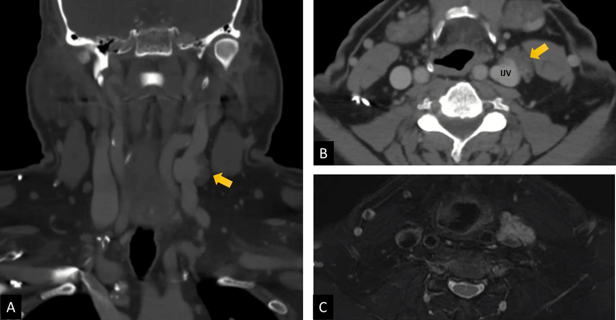
Figure 1
Preoperative imaging of internal jugular venous malformation. (A) Coronal view of computerized tomographic scan. (B) Axial view of computerized tomographic scan. (C) Magnetic resonance imaging showing hyperintensity of lesion in T2.
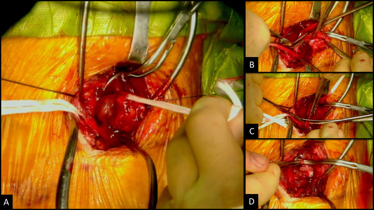
Figure 2
Surgical excision of internal jugular venous malformation. (A) Venous malformation adherent to internal jugular vein. (B) Excision of malformation. (C) Partial resection internal jugular vein. (D) Ligation of internal jugular vein stumps.
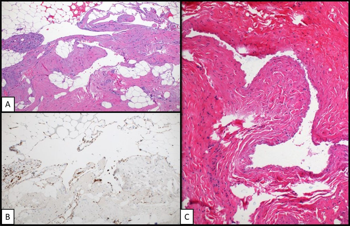
Figure 3
Histopathology analysis of resected lesion (40× magnification). (A) H&E stain of vascular malformation. (B) Immunohistochemical stain highlighting endothelial cells of vascular malformation. (C) H&E stain of left internal jugular vein.