
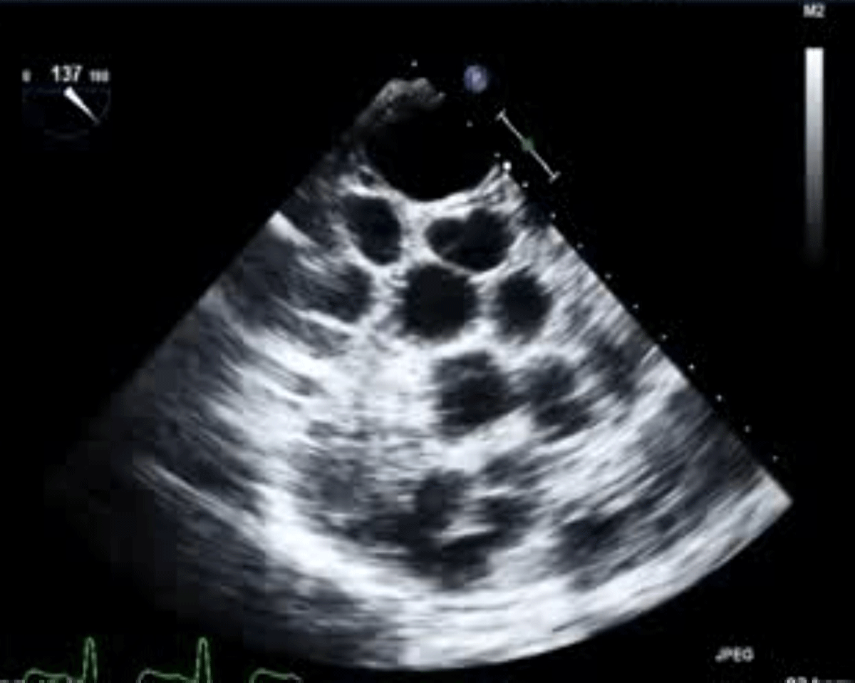
Figure 1
A transesophageal echocardiogram reveals the presence of numerous hydatid cysts entirely filling the left atrium and left ventricle cavity.
Video 1
A transesophageal echocardiogram reveals the presence of numerous hydatid cysts entirely filling the left atrium and left ventricle cavity; see also at https://youtu.be/QBTMjZTEpc8.
Video 2
Cardiac magnetic resonance imaging, cine loop. The sagittal section and short-axis view showed multiple hypointense ring-shaped thick-walled hydatid cysts occupying the entire cavity of the left atrium and left ventricle; see also at https://youtu.be/zaIBK_WH57M. LA: left atrium; LV: left ventricle
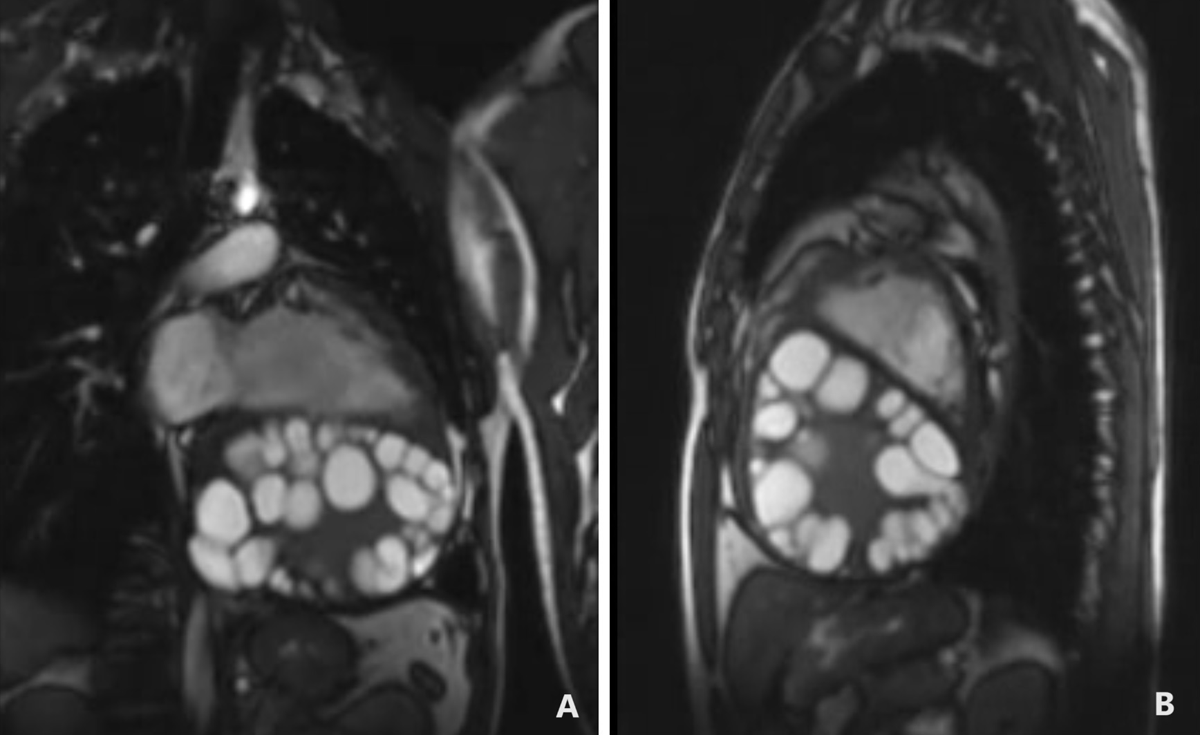
Figure 2
Cardiac magnetic resonance imaging through static images of a cine loop. (A) The sagittal section features a four-chamber view, while (B) displays a short-axis view. Both images depict multiple hypointense ring-shaped thick-walled hydatid cysts occupying the entire cavity of the left atrium and left ventricle.