
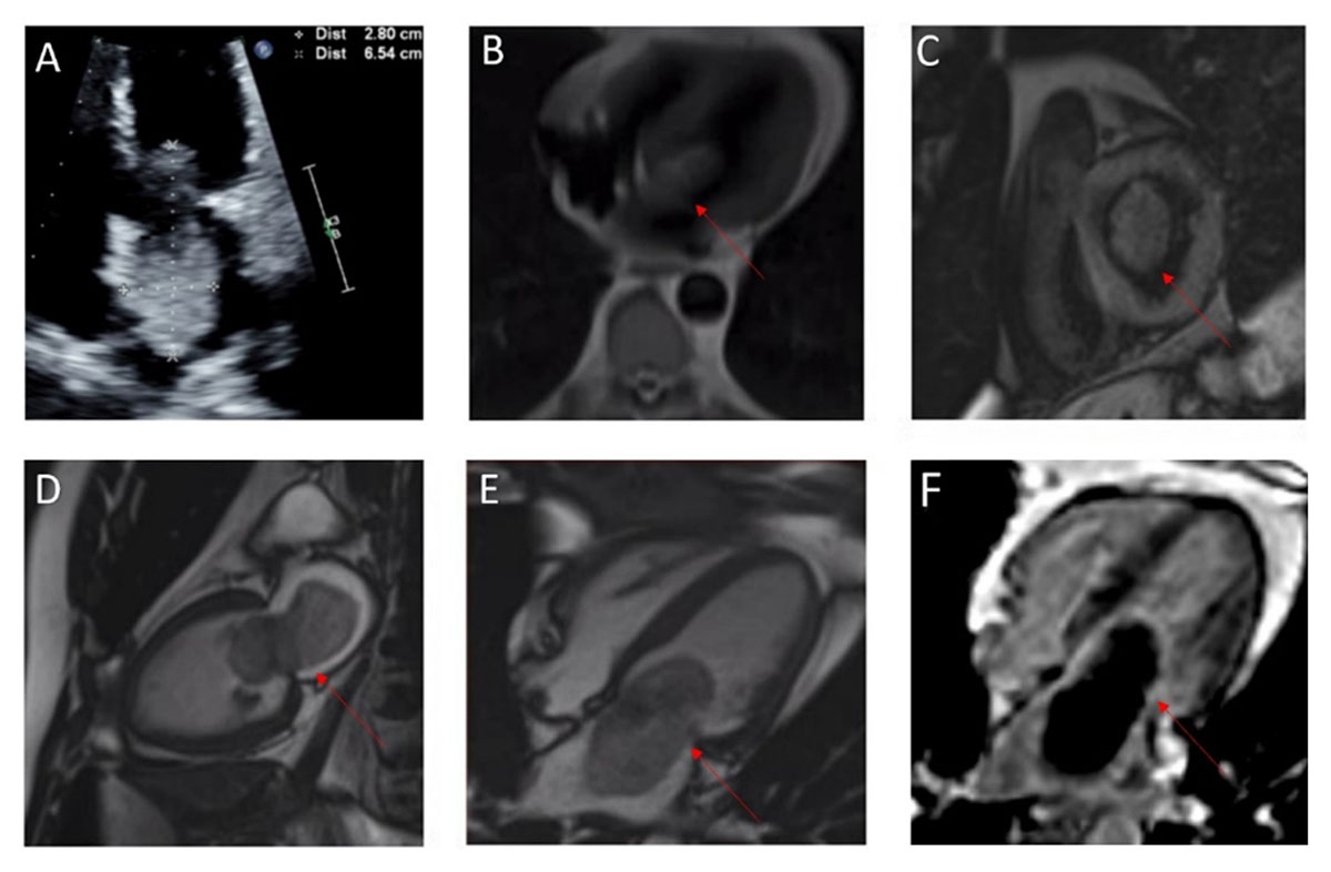
Figure 1
(A) Transthoracic echocardiogram of normal LV systolic function with an ejection fraction of 55% to 60% and large mobile echo density in the left atrium attached to the atrial septum; (B-E) cardiac magnetic resonance (CMR) imaging obtained for further characterization of the cardiac mass confirmed the presence of a large atrial mass attached to the fossa ovalis; (F) delayed gadolinium enhancement CMR with long inversion time suggested a thrombus.