
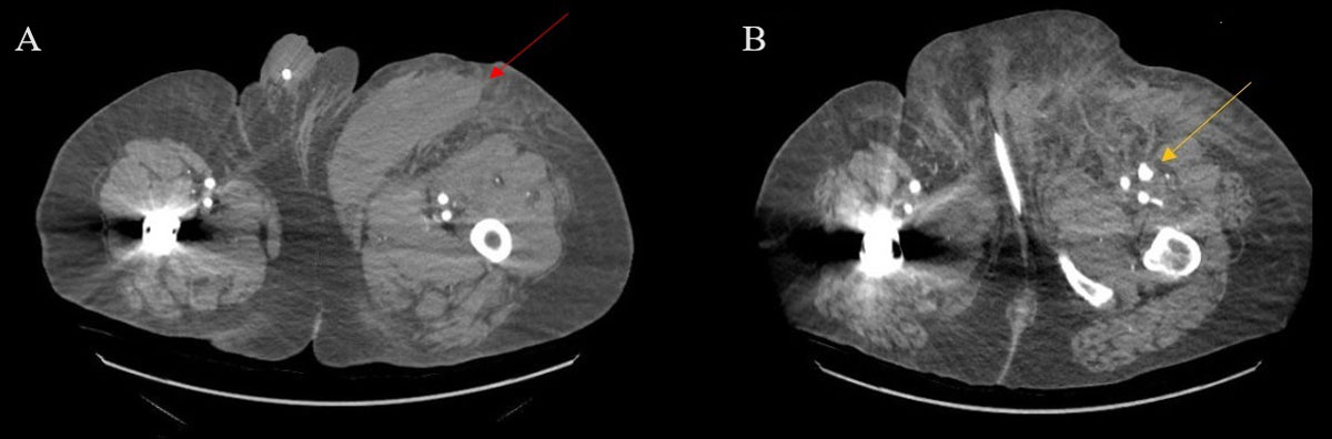
Figure 1
(A) 15 × 6 × 5.6-cm left anterior medial thigh hematoma (red arrow). (B) 3.7 × 2.1 × 1.6 cm left profunda femoral artery pseudoaneurysm with contrast extravasation (yellow arrow).
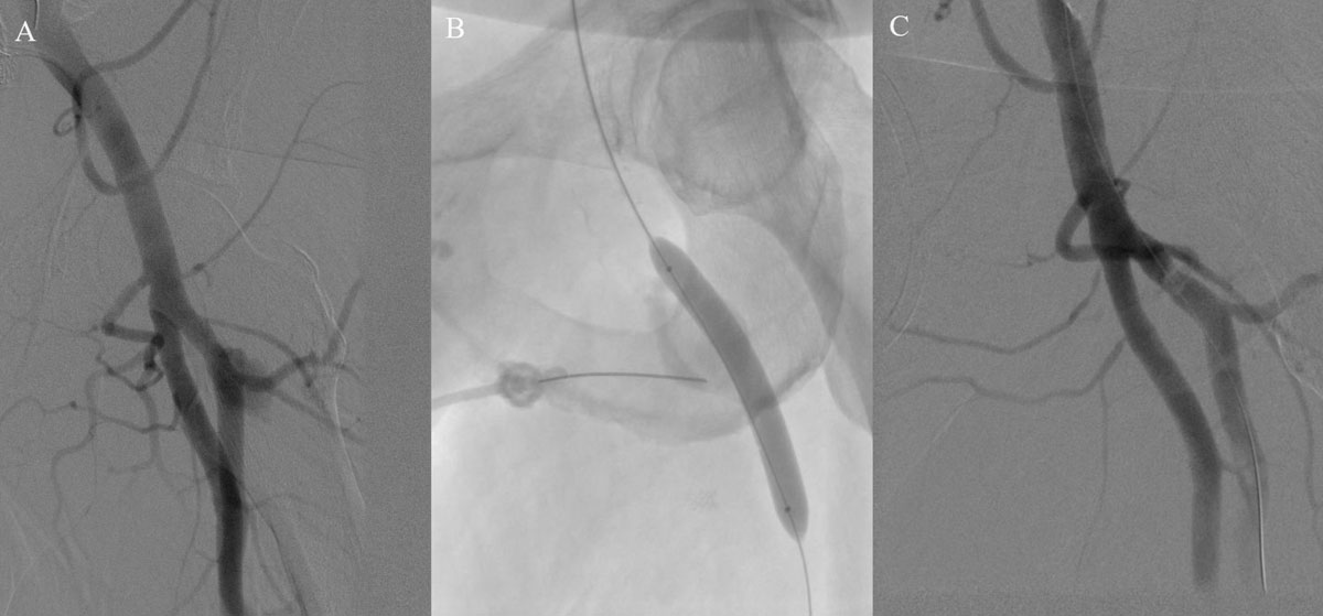
Figure 2
(A) Initial angiogram depicting the left profunda femoral artery pseudoaneurysm. (B) Imaging showing the position of the needle in the pseudoaneurysm chamber and balloon inflated in the profunda femoral artery to protect against distal embolization. (C) Final angiogram showing patency of the superficial and profunda femoral artery and resolution of the pseudoaneurysm.