
Video 1
Transesophageal echocardiogram short axis view of a pulsating left ventricular assist device (LVAD) pseudoaneurysm. Color Doppler demonstrates flow in the LVAD and at the neck of the pseudoaneurysm; see also at https://youtu.be/DSg_b3WFqbg.
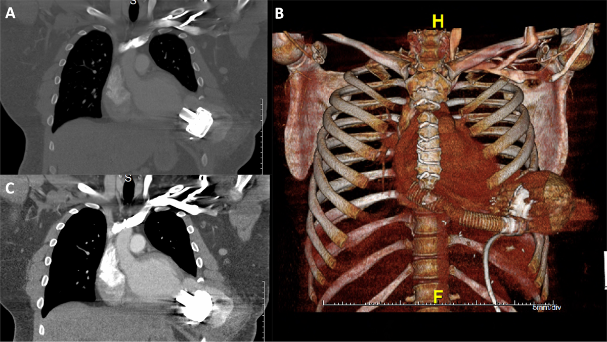
Figure 1
Computed tomography scan demonstrated a large left ventricular pseudoaneurysm measuring 10 × 10 × 8 cm in diameter surrounded by a hematoma. Coronal (A, C) and 3-dimensional reconstructed computed tomography (B) are shown. H: head; F: front