
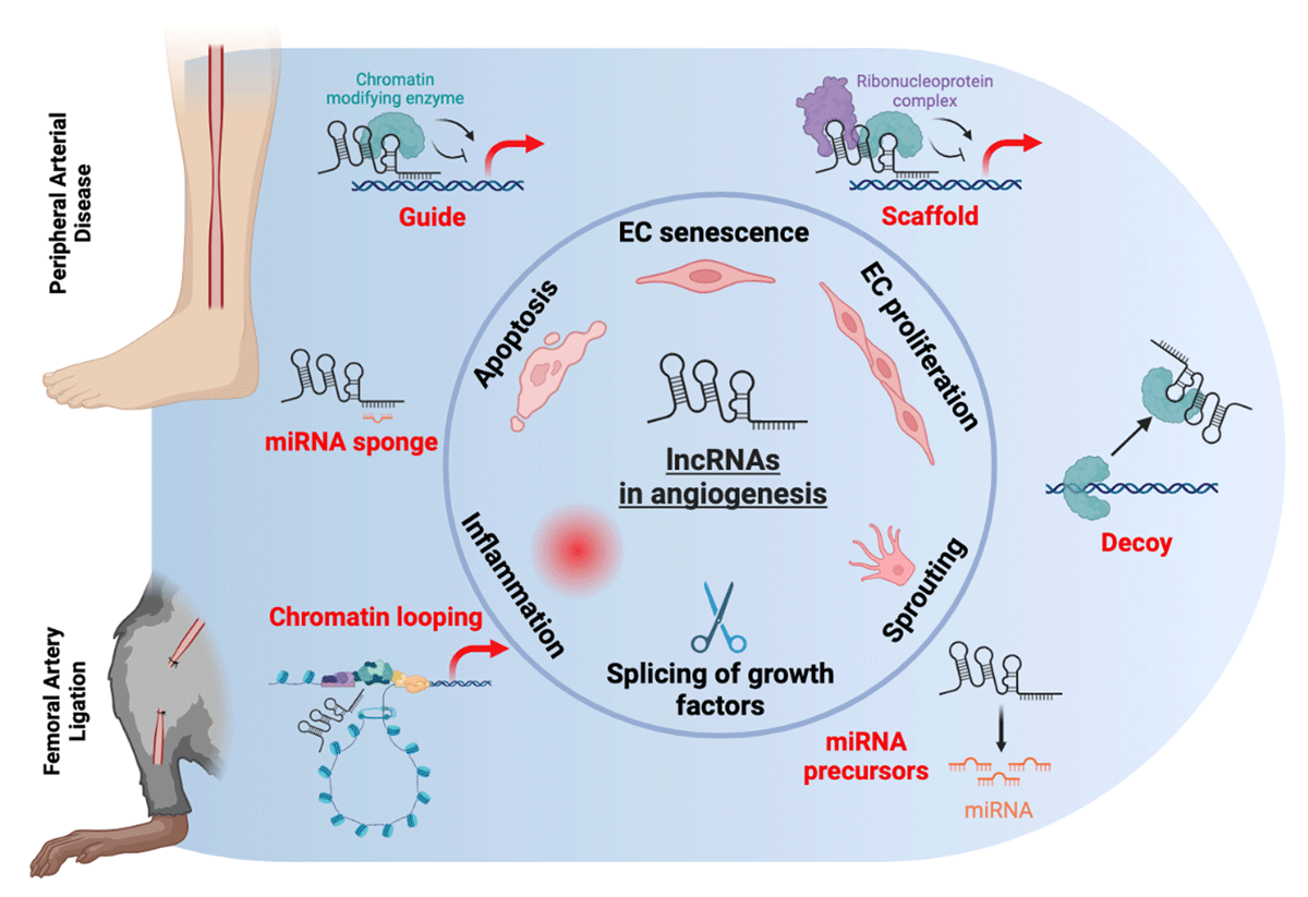
Figure 1
Potential modes of action of lncRNA in angiogenesis in peripheral arterial disease. LncRNAs are regulators of angiogenic pathways in endothelial cells, including their senescence and activation, proliferation, sprouting, and apoptosis. They also may modulate the expression of growth factors via splicing. In addition, they are involved in inflammation, a key driver of angiogenesis. The potential mechanisms through which they deploy their action are guiding, scaffolding, or decoying chromatin modifying enzymes. They also may act as miRNA sponges or precursors and are involved in chromatin structure via looping. lncRNAs: long non-coding RNAs; miRNA: microRNA
Table 1
Reported long non-coding RNAs with functional relevance in peripheral arterial disease.24,25,26,27,28,29,30,32,33,36,38,39,40,41,42,44,45,46,47,48,49,50,51,52,53,54,55,56,57,58,59,60,61,62,63 ECs: endothelial cells; KO: knockout; HLI: hindlimb ischemia; miR: microRNA; eNOS: endothelial nitric oxide synthase; KDR: kinase insert domain receptor; PBMCs: peripheral blood mononuclear cells; PAD: peripheral artery disease; VEGF: vascular endothelial growth factor; SNP: single nucleotide polymorphisms; VSMCs: vascular smooth muscle cells; BRG1: Brahma-related gene 1
| LNCRNA | CELL SPECIFICITY | RELATIONSHIP TO PAD | PROPOSED MECHANISM |
|---|---|---|---|
| MALAT124,25,26,27,28 | Multiple cell types including ECs | Reduced blood flow recovery in KO model of HLI, involved in EC proliferation | VEGFR2 |
| MEG329,30,32,33 | Multiple cell types including ECs and fibroblasts | Improved blood flow recovery in KO model of HLI, expressed in senescent ECs and reduced during EC sprouting | Notch signaling, miR-21 |
| ANRIL36,38,39,40,41,42 | Multiple cell types including ECs, myocytes and fibroblasts | cdkn2b-deficient mouse under HLI showed reduced blood flow recovery and higher digital amputation | TGF-βR1/Smad pathway, eNOS |
| SNHG1244,45 | Multiple cell types including ECs, immune cells and fibroblasts | In KO mice, impaired angiogenic response to HLI, more pronounced in diabetes | Wnt, Notch, and angiopoietin signaling pathways |
| LEENE46,47,48,49 | EC enriched | Impaired perfusion recovery in HLI of KO mice, restored with LEENE expression | KDR, eNOS |
| STEEL50,51,52 | Endothelial enriched | Stimulates formation and maturation of vascular flow networks; decreased expression in disturbed flow | KD reduces expression of shear stress related genes KLF2, eNOS; feedback loop with KLF2 |
| H1952,53,54 | Multiple cell types including ECs, myoblasts and PBMCs | Increased in PAD mouse model after 14 days of ischemia, involved in myogenesis, reduced capillary density EC specific-KO model of HLI | STAT3 signaling; IL-6; VEGF |
| MIR22HG52,54 | Multiple cell types including ECs | Elevated in hypoxic ECs and HLI model | Unknown |
| LINC0060755,56,57 | ECs and VSMCs | Associated SNP found predisposing patients to PAD | c-Myc |
| HIF1A-AS158,59 | VSMCs and ECs | Hypoxia regulated element | BRG1 |
| SENCR52,60 | Highest in ECs but also expressed in VSMCs | Expression is reduced in human critical limb ischemia patients | CX1CL3, CCL5, CEACAM1 |
| FENDRR61 | ECs and VSMCs | Decreased in hypoxia | DRP1 DNA methylation, p53 |
| HIF1A-AS262,63 | VSMCs and ECs | Hypoxia regulated element | USF1 to elevate ATF2 |
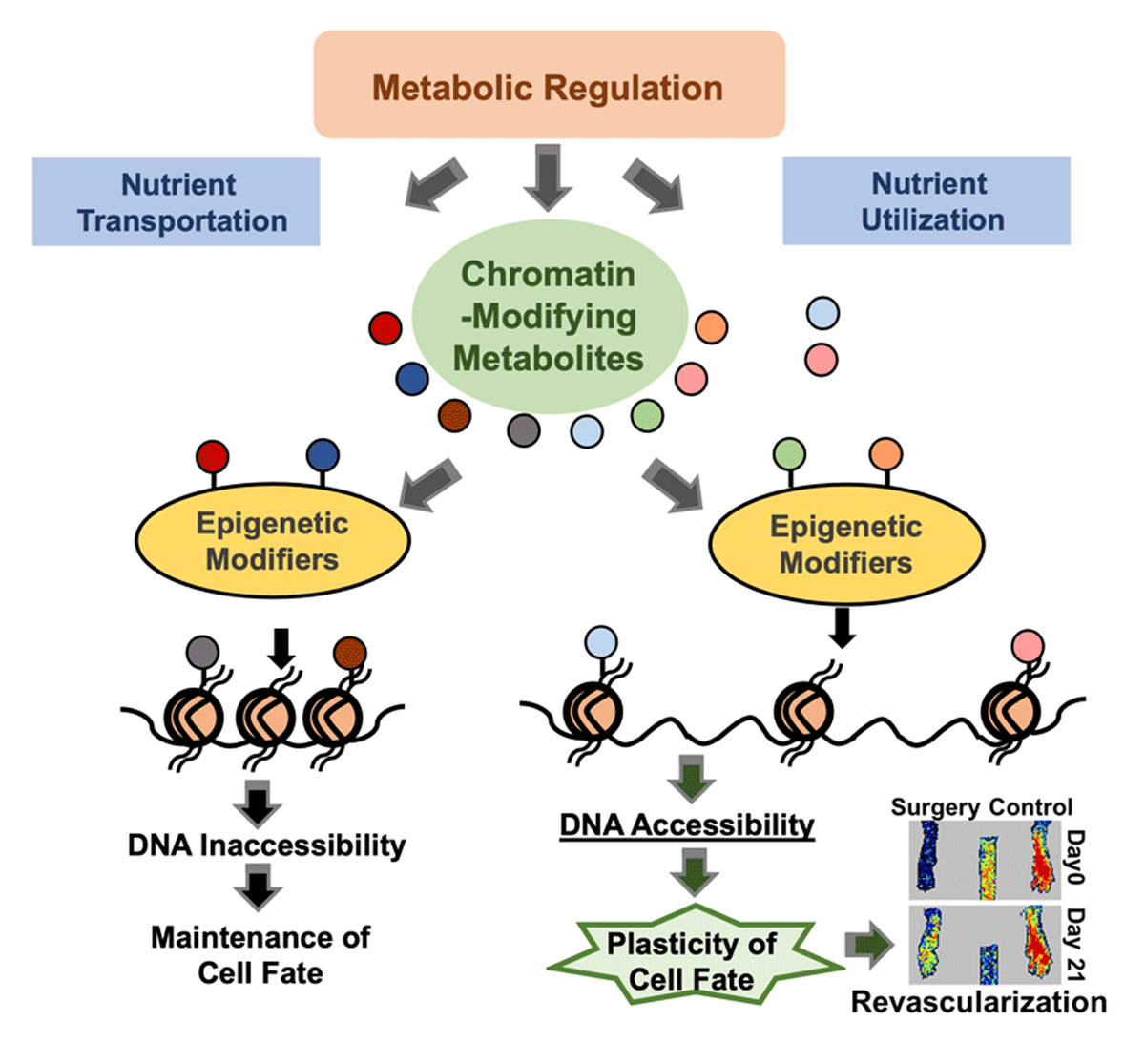
Figure 2
Metabolic regulation on epigenetics. The chromatin-modifying metabolites, regulated by their rate-limiting enzymes and the availability of their precursors, can bind to histone tails or epigenetic modifiers to affect chromatin accessibility, which further modulates the cell fate transition.