
Video 1
Subcostal echocardiography view showing a rounded structure measuring 6.8 cm in diameter adjacent to and slightly compressing the right atrium; see also at https://youtube.com/shorts/k97OoTMpAbo.
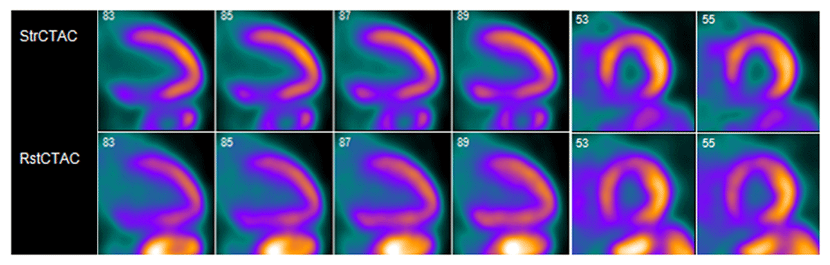
Figure 1
Cardiac positron emission tomography showing a medium-sized, moderate-severity, partially reversible defect in the proximal and mid inferior and inferoseptal segments.
Video 2
Coronary angiogram showing an ectatic left coronary system and an incompletely opacified ectatic proximal right coronary artery; see also at https://youtube.com/shorts/0ssjghqx5sM.
Video 3
Cardiac computed tomography angiography sagittal view showing a giant retrosternal right coronary artery aneurysm extending from the proximal to distal segment, compressing the right atrium; see also at https://youtu.be/HMw3lXG_90c.
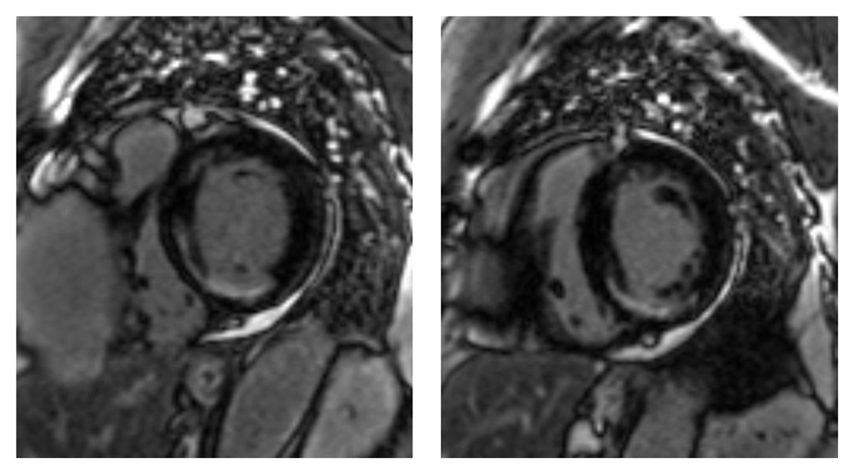
Figure 2
Late gadolinium enhancement cardiac magnetic resonance imaging showing left ventricular subendocardial scar in the basal and mid inferior and inferoseptal walls consistent with right coronary artery related myocardial infarction.