
Figure 1
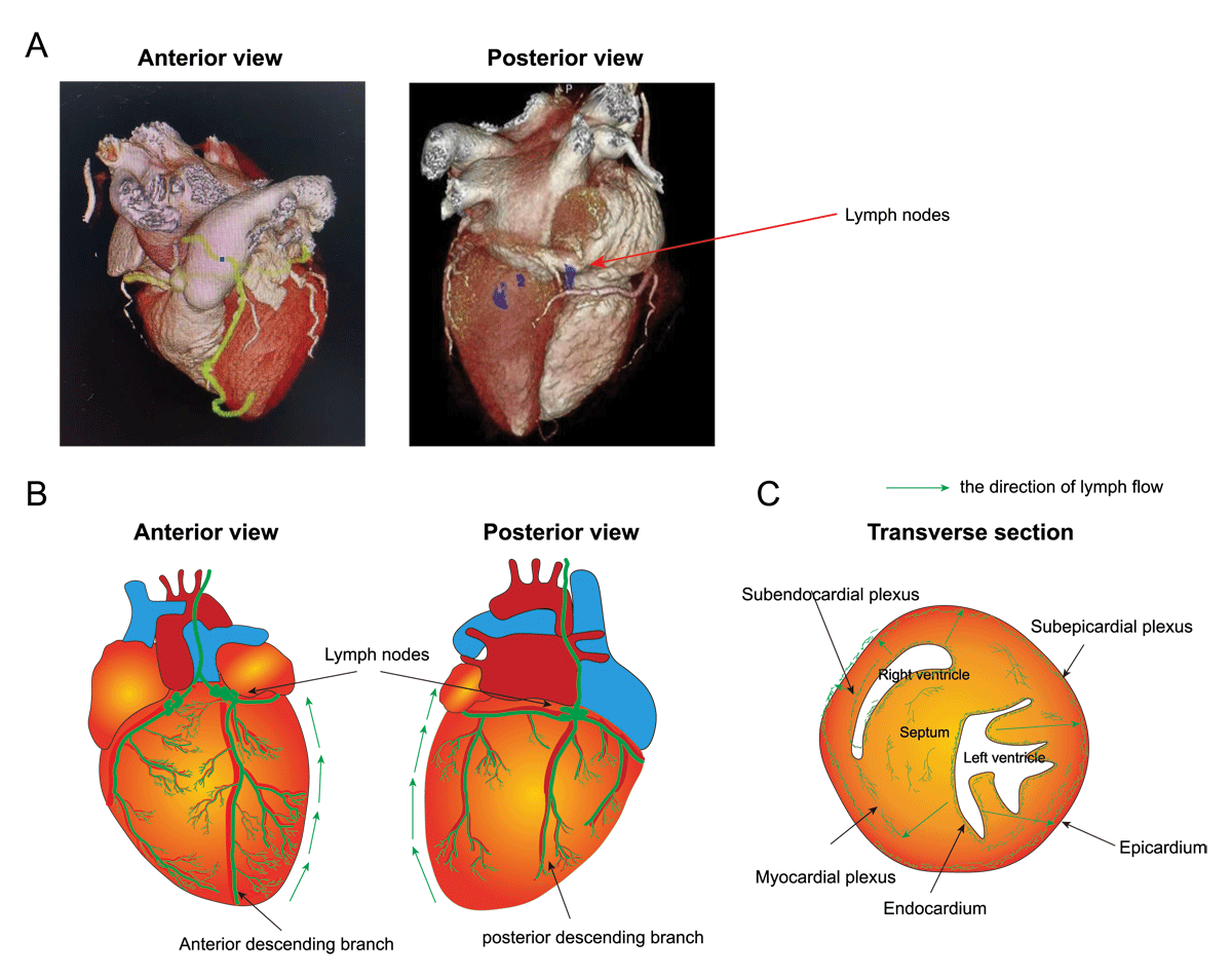
Structure and function of cardiac lymphatic vessels. (A) Three-dimensional reconstruction after cardiac computed tomography (CT) scanning showing both anterior and posterior view of the human heart. The red arrow indicates lymph nodes. (B) Illustration showing the distribution pattern of cardiac lymphatic system in the human heart. (C) Transverse section of the heart showing subendocardial, subepicardial, and myocardial plexus of cardiac lymphatics.
Table 1
Models of cardiovascular diseases.
| DISEASE MODEL | APPROACH TO PROMOTE LYMPHATIC GROWTH | EFFECTS | REFERENCES |
|---|---|---|---|
| MI | VEGF-C | Promotes lymphangiogenesis, reduces myocardial edema, alleviates the degree of inflammation and fibrosis, and improves cardiac function | 11, 23, 32 |
| Ephrinb2 signaling | Enhances lymphangiogenesis | Data unpublished | |
| Adrenomedullin, Apelin, NTS- and Reelin | Drives repair cardiac lymphangiogenesis and function | 33-36 | |
| Hypertension and dermal lymphangiogenesis | VEGF-C | Activates local lymphangiogenesis | 43-44 |
| A2AR | Increases lymphatic capillary density and sodium clearance in the skin and reduces blood pressure | 46 | |
| Infective endocarditis | VEGF-A | Induces lymphatic vessel enlargement | 49 |
| VEGF-C | Induced sprouting lymphangiogenesis | 52 | |
| Cardiac transplantation | Inhibition of VEGF-C/VEGFR-3 | Reduces early lymphoid activation, subsequent acute and chronic inflammatory responses | 61 |
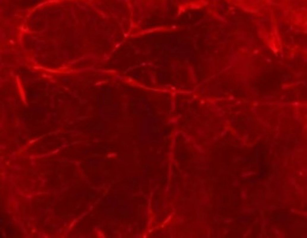
Figure 2
Visualization of endogenous tdTomato (red) fluorescence of hearts from Lyve1-Cre; Rosa26-tdTomato mice. Scar bar: 1 mm.