
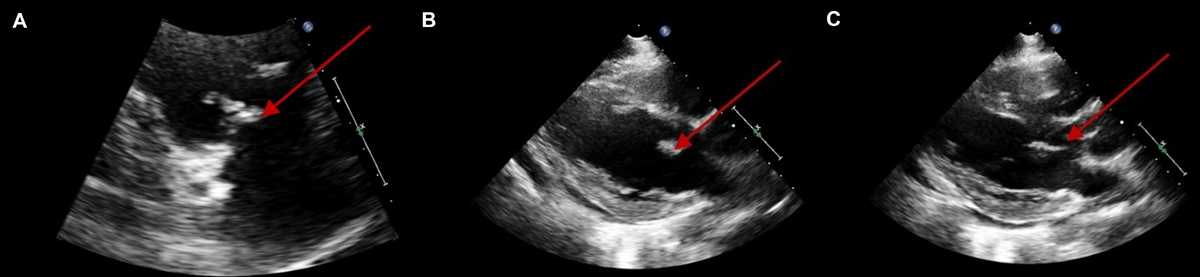
Figure 1
Transthoracic echocardiography of trivalvular vegetation. Vegetations observed on transesophageal echocardiography were as follows: (A) tricuspid valve (16 × 8.1 mm), (B) mitral valve (13.6 × 8.0 mm), and (C) aortic valve (18.3 × 4.7 mm).
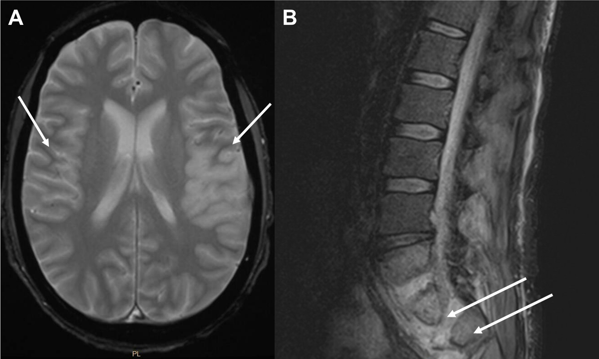
Figure 2
Brain and spinal cord infarctions and abscess. (A) Magnetic resonance (MRI) of brain with and without (w/wo) contrast with left and right middle cerebral artery infracts and (B) L spine MRI showing L5-S1 discitis-osteomyelitis with intramuscular paraspinal abscess at the L5 and S1 levels.