
Figure 1
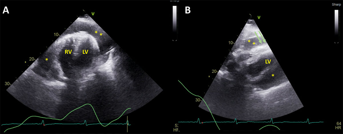
Transthoracic echocardiography, apical 4-chamber (A) and subcostal view (B) showing large circumferential pericardial effusion (asterisks) with right ventricular diastolic collapse (green arrows). LV: left ventricle; RV: right ventricle
Video 1
Transthoracic echocardiography with Doppler showing hemodynamically significant large pericardial effusion with right-sided chamber collapse and respiratory variation across the inflow; see also at https://youtu.be/VI1MBbjn2Vw.
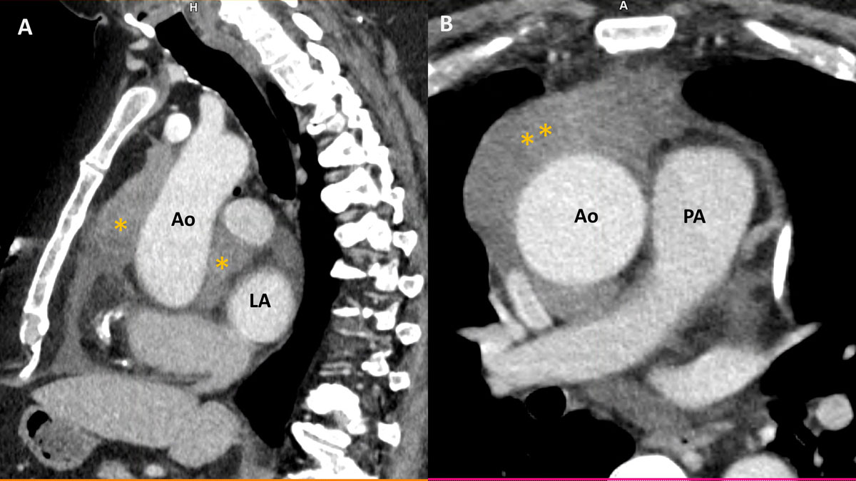
Figure 2
Contrast-enhanced computed tomography scan of the chest, sagittal view (A), and axial view (B) showing a hyperintense (Housefield unit of 50) large lobulated anterior mediastinal mass (asterisks) encasing the aorta and extending posteriorly to the left atrium. Ao: aorta; LA: left atrium; PA: pulmonary artery
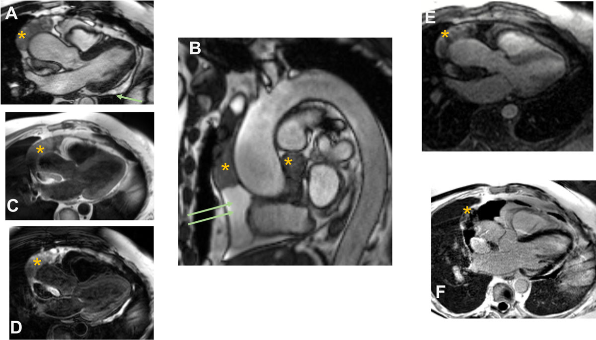
Figure 3
Cine cardiac magnetic resonance imaging. (A) 3-chamber view showing heterogenous lobulated intrapericardial mass (asterisk) with a posterior pericardial effusion (arrow). (B) Sagittal candy-cane view of the aorta showing pericardial effusion (arrows) and masses (asterisk). (C-D) T1- and T2-weighted imaging showing isointensity on T1 and hyperintensity on T2. (E-F) First pass perfusion and late gadolinium imaging showing evidence of contrast uptake on the first pass and heterogenous signal intensity suggesting patchy late gadolinium uptake.
Video 2
Noncontrast, axial bright blood imaging showing large heterogenous intrapericardial mass extending superiorly to the arch and encasing the aorta; see also at https://youtu.be/pnJ-g_lxM0c.
Video 3
First pass perfusion with cardiac magnetic resonance showing contrast uptake; see also at https://youtu.be/3qS6IYbRYxk.
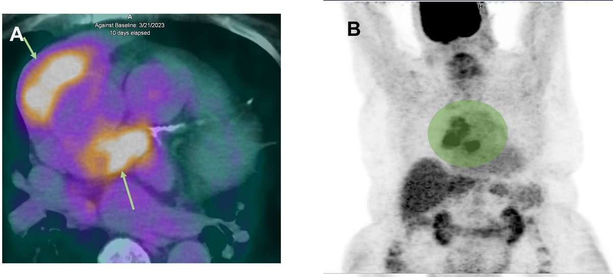
Figure 4
(A) Fluorine-18-fluorodeoxyglucose (18F-FDG) positron emission tomography/computed tomography shows positive FDG uptakes in periaortic pericardial masses (arrows). (B) Corresponding whole body PET/CT imaging with green area showing increased metabolic activity and FDG uptake but no evidence of mediastinal lymph node or extracardiac metastasis.