
Introduction
Variations in blood pressure above or below the physiological baseline is a common occurrence in the critical care unit, particularly in patients with underlying cardiovascular pathology. The distinction between hypotension and hypoperfusion is particularly critical in the cardiac intensive care unit (CICU), specifically for patients in the perioperative period. The perfusion of an organ is dependent on the mean arterial pressure, which is a function of cardiac output and systemic vascular resistance. A shift in thought process around the definition of shock may be the presence of hypoperfusion, even in the setting of normotension, which is generally thought of as “pre-shock” or compensated shock. Unsurprisingly, outcomes, including mortality, are worse in patients with evidence of both hypotension and hypoperfusion.1 In this review, we aim to identify appropriate blood pressure targets in the ICU, recognizing special patient populations and outlining the risk factors and predictors of end organ failure.
Appropriate Blood Pressure Target in a Critically Ill Patient
Historically, mean arterial pressure (MAP) has defined blood pressure target thresholds in the critical care unit. Every unit of time-weighted average (TWA) spent at MAP < 65 mm Hg has been strongly associated with myocardial infarction, acute kidney injury, and mortality in the critical care unit.2 This goal MAP is recommended as the lowest pressure to target initial resuscitation in patients with septic shock by the guidelines of the Surviving Sepsis Campaign.3 The extrapolation of the same threshold to all patient types, pathologies, and comorbidities is a challenge and certainly deserves more investigation.
Randomized trials of blood pressure targets are inherently difficult. Asfar and colleagues performed the largest such trial in septic shock patients and compared mean pressures of 80 mm Hg to 85 mm Hg (high target group) with current recommendations of 65 to 70 mm Hg (low target group) in patients with septic shock undergoing resuscitation. The primary outcome was mortality at 28 and 90 days, and they found no difference.4 Importantly, pressures were about 5 mm Hg higher in each group, and this was likely a trial of two higher pressures, albeit with adequate intergroup separation.
A recent retrospective analysis from a well-validated electronic ICU dataset with nearly 80,000 patients found that components of blood pressure, namely mean, systolic, and diastolic pressures, were comparable in their strength of association with organ system injury and ICU mortality.5 Estimated change-points for the risk of ICU mortality in septic patients were 69 mm Hg for mean, 100 mm Hg for systolic, 60 mm Hg for diastolic, and 57 mm Hg for pulse pressure.5 Other observational data derived from large datasets show that higher thresholds of MAP closer to 80 to 85 mm Hg may be the inflection point for increasing harm in both the medical and surgical ICU population.2,6,7 While it is difficult to separate intraoperative hypotension from postoperative hypotension in the ICU, even previously normotensive operating room patients suffer harm with new-onset hypotension in the critical care unit.8 Thresholds at 75 mm Hg or higher may be associated with decreased delirium and improved arousal levels as measured by sedation scales.7,9 In the SEPSISPAM (Sepsis and Mean Arterial Pressure) trial, patients with chronic hypertension had significantly more renal injury if they were exposed to a MAP of 70 to 75 mm Hg (ie, in the lower target group) while more rhythm abnormalities occurred in the higher target group at a MAP of 85 to 90 mm Hg.4 Lamontagne and colleagues in the 65-trial showed no difference in survival when patients were randomized to a MAP of at least 65 mm Hg versus permissive hypotension at a MAP of 60 mm Hg.10
In the critically ill cardiac patient population, diastolic blood pressure (DBP) is an important determinant of coronary blood flow, and low DBP, particularly < 60 mm Hg, has been associated with increased risk of myocardial injury.11 Critically ill patients with cardiogenic shock and a 24-hour average MAP < 65 mm Hg have a rapidly increasing rate of mortality, exceeding 70% with a 24-hour average MAP < 60 mm Hg.12 Further, patients with sustained (duration > 10 minutes), MAP < 64 mm Hg before, during, and after cardiopulmonary bypass (CPB), particularly those with sustained MAP < 55 mm Hg, have a strong association with acute perioperative stroke.13 Other investigators have confirmed that duration of hypotension per 10 minutes of a MAP < 65 mm Hg during an entire cardiac surgery and after CBP is equally important.14
The conundrum, therefore, is whether we should target higher pressures in all patients or in more vulnerable ones. Furthermore, it seems that the mechanisms to achieve higher pressures may be as important as the goal itself. Exposure to supratherapeutic doses of catecholamines may not be desirable.15,16 Almost 10 years after the impressive work by the SEPSISPAM group, we need to think of trials with appropriate patient populations, targets, and outcomes. Certainly, the concept of perfusion pressure being the difference of MAP and central venous pressure or in some cases intra-abdominal pressure is a critically important area of focus.17 Perfusion pressure may be extrapolated to renal perfusion and a necessary threshold of at minimum 60 mm Hg. Analysis comparing perfusion pressure deficits and MAP deficits has been seen to have a nearly similar and strong association with ICU mortality at two weeks.18,19
Cardiac Critical Care: Different Patient Populations and Personalized Blood Pressure Targets
Individualization of MAP goals is important in the post-cardiac surgery patient, as different comorbidities, operative interventions, and the presence of mechanical circulatory support, among other criteria, may influence the MAP required to maintain adequate perfusion (Table 1).
Table 1
Individualized blood pressure management in the post-cardiac surgery patient. SAM: septal anterior motion; CPB: cardiopulmonary bypass; MAP: mean arterial pressure; HCOM/SAM: hypertrophic obstructive cardiomyopathy/systolic anterior motion; LVOT: left ventricular outflow tract; LVH: left ventricular hypertrophy; SBP: systolic blood pressure; VSD: ventricular septal defect; LVAD: left ventricular assist device; VV vs VA ECMO: venovenous vs venoarterial extracorporeal membrane
| A. Decision points for individualized blood pressure targets for post cardiac surgery patients |
| 1. Age |
| 2. Nature of surgery |
| 3. Intraoperative course and intraoperative data |
| 4. Left ventricular versus right ventricular dysfunction |
| 5. Presence or absence of mechanical circulatory support devices |
| B. Patients who need a higher mean arterial pressure goal to optimize tissue perfusion |
| 1. Right ventricular (RV) failure |
| 2. Post-heart transplant with RV dysfunction |
| 3. Hemodynamic instability on weaning from CPB |
| 4. Air in the coronary arteries post-cardiac surgery needing higher MAP goals during initial few hours postoperatively |
| 5. Patients with long CPB time and cross-clamp time |
| 6. Acute kidney injury identified by low urine output intraoperatively |
| 7. HCOM/SAM needing higher MAP to stent open the LVOT |
| 8. Patient with LVH |
| 9. History of chronic hypertension with shift in cerebral autoregulation |
| C. Patients who need a lower MAP goal to optimize perfusion |
| 1. Patients with LV dysfunction where a reduction in afterload will help increase cardiac output |
| 2. Post aortic surgery, complex congenital repairs, fistula repairs and VSD repairs where a higher-than-normal SBP/MAP can cause increased tension on critical suture sites and lead to catastrophic bleeding |
| 3. Patients with circulatory support devices LVAD, impella, VV vs VA ECMO where increase in systemic arterial blood pressure can compromise flow through these support devices |
Most commonly, patients with chronic uncontrolled hypertension are presumed to have a rightward shift in their cerebral autoregulation curve, thereby requiring a higher average MAP goal to maintain cerebral blood flow and perfusion. A frequently referenced threshold for these patients is a minimum MAP no less than 20% from their baseline blood pressure, usually averaged over several previous measurements.20 Mechanistically, a worsened microcirculation in the chronically hypertensive and the need for a higher perfusion pressure to maintain end-organ perfusion contribute to the underlying pathophysiology.21
After post-acute myocardial infarction, higher MAP goals are associated with smaller infarct sizes, and thus use of vasopressor and inotropic support to obtain higher mean pressures is recommended in this population.22
Patients with uncorrected valvular lesions and physiology that requires higher afterload may also require higher MAP goals. These specifically include patients with clinically significant aortic stenosis, left ventricular (LV) hypertrophy, and those with septal anterior motion of the anterior leaflet of the mitral valve due to hypertrophic obstructive cardiomyopathy or asymmetric septal hypertrophy. Finally, patients who develop tamponade physiology postoperatively require higher afterload until the pericardium can be decompressed. Given ventricular interdependence, maintenance of normal to slightly increased systemic blood pressure is critical to maintaining the transseptal pressure gradient and thus normal wall motion of both the left and right ventricles. Consequently, those with significant right ventricular dysfunction, including those post-orthotopic heart transplantation, will require higher MAP goals.23
Other specific populations that may benefit from higher MAP goals include patients with coronary air embolism during weaning from CPB,24 patients with a prolonged CPB run or cross-clamp time, and those who have difficulty weaning from CPB, which may be associated with relative coronary ischemia. Finally, poor perioperative urine output may indicate end-organ hypoperfusion and increased risk of acute kidney injury, so such patients may benefit from higher perfusion pressure.25 In contrast, patients with hemodynamically significant aortic insufficiency and mitral regurgitation require reduced afterload. Lower MAP goals within limits and without compromising on coronary or another end-organ perfusion may be necessary.
Aside from patients with valvular or other pathology, a small subset of patients may have indications for lower MAP goals. Predominantly, this group includes patients who have undergone aortic surgery, those with complex congenital repairs (including fistulas), and those with ventricular septal defects—all cases in which increased MAP leads to tension on critical suture lines, causing possible disruption that can lead to catastrophic and potentially irreparable bleeding. However, recent emerging evidence suggests that this may be an exaggerated association of lower clinical significance.26
Another subset of patients in whom decreased systemic blood pressure may be appropriate is those with mechanical circulatory support devices, including temporary and durable LV assist devices, as well as extracorporeal membranous oxygenation through which flow may be compromised in high afterload states.21 Appropriate MAP targets in this setting remain a particular enigma since increasing pressure with afterload most surely compromises forward flow in most of these patients.
If relative hypotension is deemed appropriate in a specific patient, close monitoring of end-organ function is mandatory, and indications should be repeatedly addressed to liberalize toward normal MAP goals as soon as it is safe.
Choosing Pressure versus Indicators of Tissue Perfusion when Managing Shock States
While initial presentation of shock includes evidence of hypoperfusion and hypotension, not all such states are equal (Figure 1).27 Cardiogenic shock (CS) is a continuum of hemodynamic instability, including isolated hypotension with preserved organ perfusion, isolated organ hypoperfusion despite preserved blood pressure, and the combination of hypotension and hypoperfusion.1 Figure 2 shows the Society for Cardiovascular Angiography & Interventions classification of CS, which aims to provide a standardized classification system across multidisciplinary teams to help provide consistent care and prognostic utility to these high-risk critically ill patients. However, further validation studies are required to determine whether the SCAI classification adds significantly to prognostication and outcomes.28
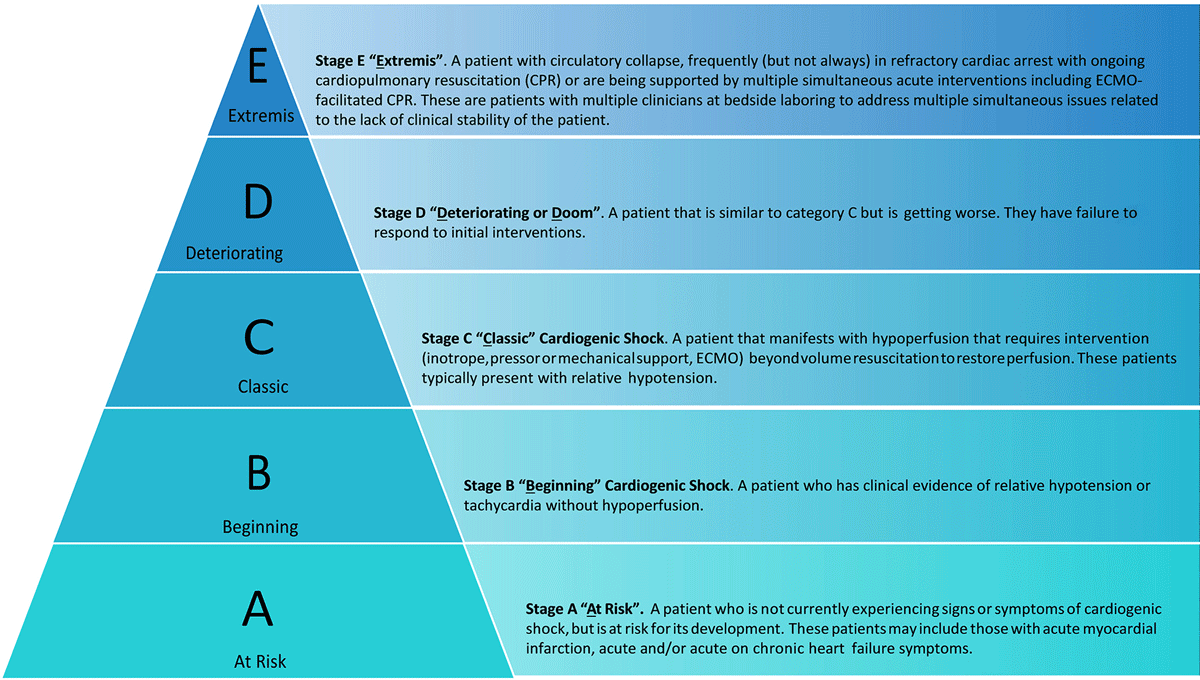
Figure 1
Society of Cardiovascular Angiography and Interventions (SCAI) classification of cardiogenic shock. Reprinted with permission.28 ECMO: extracorporeal membrane oxygenation; CPR: cardiopulmonary resuscitation
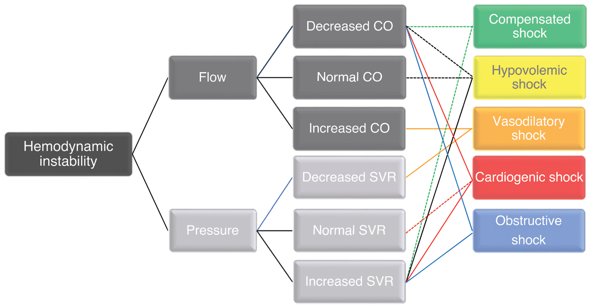
Figure 2
Effects of cardiac output and afterload and the inter-relationship with end-organ perfusion. CO: cardiac output; SVR: systemic vascular resistance
Hypotension and hypoperfusion are both associated with increased mortality in CICU patients. Hospital mortality is higher with isolated hypoperfusion or concomitant hypotension and hypoperfusion (classic shock); therefore, patients with hypoperfusion can be considered to have shock irrespective of blood pressure.1 However, the equation of MAP with markers of tissue perfusion and microcirculation per se is still up for debate.29 Capillary microvascular density and flow at high dose vasopressors to maintain MAP compared to normotension with no vasopressors is very different. Capillary refill time has been the subject of recent exploration and was found to be no different from lactate in guiding resuscitation in septic shock.30 A recent meta-analysis reported a very weak negative correlation between capillary refill time and MAP.31
The specific effects on flow and pressure can vary depending on the underlying cause and severity of the shock. In general, CS is characterized by a decrease in cardiac output, which can lead to a decrease in blood pressure. However, the relationship between flow and pressure in CS can be complex as systemic vascular resistance increases to compensate for the decreased cardiac output; this can help maintain blood pressure but may further decrease cardiac output. The goal is to optimize cardiac output and tissue perfusion by targeting a specific flow in addition to blood pressure (Figure 3). This can be assessed by measuring cardiac output directly using a pulmonary artery catheter, a minimally invasive tool that uses arterial waveform analysis with or without surface or transesophageal echocardiography (TEE) and in conjunction with trending markers of tissue hypoperfusion (Table 2).
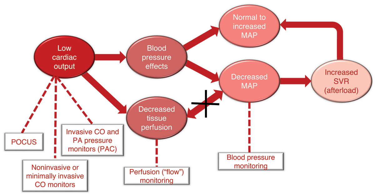
Figure 3
Cardiogenic shock with hypotension and hypoperfusion and compensated shock with normal or increased afterload. POCUS: point of care ultrasound; CO: cardiac output; MAP: mean arterial pressure; SVR: systemic vascular resistance; PAC: pulmonary artery catheter
Table 2
Clinical, diagnostic, and laboratory markers of tissue hypoperfusion.
| 1. Decreased blood pressure: systolic < 90 mm Hg OR mean pressure < 60 mm Hg OR > 30 mm Hg drop from baseline |
| 2. Tachycardia: heart rate > 100 beats/min |
| 3. Weak or absent peripheral pulses |
| 4. Cool, clammy, or mottled skin |
| 5. Altered mental status |
| 6. Decreased urine output < 30 mL/hr |
| 7. Cardiac index < 2.2 |
| 8. Pulmonary capillary wedge pressure > 15 mm Hg |
| 9. Right atrial pressure ≥ 0.8 mm Hg |
| 10. Pulmonary artery pulsatility index < 1.85 |
| 11. Cardiac power output ≤ 0.6 watts |
| 12. Mixed venous oxygen saturation (SvO2) < 60% |
| 13. Elevated lactate > 2 mmol/L |
| 14. Creatinine doubling |
| 15. 50% drop in glomerular filtration rate |
| 16. Deranged liver function |
| 17. Elevated NT-pro brain natriuretic peptide |
Monitoring and Diagnostic Modalities to Guide Blood Pressure Management in the ICU
Arterial blood pressure is determined primarily by cardiac output, systemic vascular resistance, vascular compliance, and blood volume.32 In the ICU, and especially in the cardiac critical care unit, use of an “invasive continuous arterial blood measurement” is widely considered the gold standard. However, blood pressure alone is simply not enough in these patients. Therefore we are seeing a consistent move away from static toward dynamic measurements.33 Beyond knowing that a patient is hypotensive, a critical care physician needs to determine whether the patient is fluid responsive or needs vasopressor or inotropic support. To this end, dynamic variables such as plethysmography variability index, pulse pressure variation, and stroke volume variation are helpful and increasingly used as easy bedside tools.33
Plethysmography Variability Index
Plethysmography variability index (PVI) utilizes changes in pulse oximetry waveforms over the entire respiratory cycle to predict fluid responsiveness.33 In a prospective observational study of mechanically ventilated patients undergoing noncardiac surgery, PVI was measured and a fluid bolus given. Patients with a PVI > 10.5% were noted to have a decrease in heart rate with concurrent increase in cardiac index following the bolus.34 Forget et al. likewise showed that during major abdominal surgery, use of PVI to guide fluid therapies led to decreased lactate levels when compared with standard care both during surgery and 48 hours postoperatively.35
Pulse Pressure Variation
Pulse pressure variation (PPV) has been a popular marker of fluid responsiveness since it requires no additional equipment beyond an invasive blood pressure monitor. To be accurate, it does require appropriately large tidal volumes in a mechanically ventilated patient using volume control ventilation in normal sinus rhythm. Yang and Du undertook a recent meta-analysis to evaluate PPV in the critical care population.36 This examination of 22 studies found PPV had a pooled sensitivity of 0.88 and pooled specificity of 0.89 for fluid responsiveness.36
Stroke Volume Variation
Stroke volume variation (SVV) can be evaluated by one of several commercially available monitors that differ in their method of estimating aortic impedance.37 In a meta-analysis of 24 studies utilizing SVV in the surgical and critical care population, Zhang et al. found SVV highly correlated with fluid responsiveness (r = 0.72) and with a sensitivity and specificity of 0.81 and 0.80, respectively.37 The correlation was found to be even stronger in the ICU population, which the authors postulated was due to the relative hypovolemic state of these patients compared with those in the operating room when evaluated.37
In addition, an important and easy to use bedside tool in the ICU is the tidal volume challenge that uses the change of tidal volume from the typical lung protective 6 mL/kg to a larger target of 8 mL/kg. This change in SVV and PPV obtained by transiently increasing tidal volume (tidal volume challenge from 6 to 8 mL/kg) was superior to PPV and SVV in predicting fluid responsiveness during low tidal volume ventilation. Myatra and colleagues demonstrated this in a study in 20 patients in 2017, and these findings were validated in various larger external cohorts in subsequent analyses.38,39
Echocardiography
The important role of echocardiography in the critical care setting cannot be overstated. Regardless of modality, echocardiography can be a useful tool for examining hemodynamic failure, assessing etiology of cardiopulmonary arrest, and determining volume status via superior vena cava (with transgastric TEE) or inferior vena cava (with transthoracic echocardiogram) collapsibility.40 In an extensive review of the literature, Prager et al. noted that utilization of TEE in the critical care setting resulted in a change of diagnosis in 52% to 78% of patients and a change in management in 32% to 79% of patients examined.41 They additionally showed that TEE was able to successfully distinguish the etiology of cardiac arrest in 25% to 35% of cases when it was used for this purpose.41
As shown in Figures 4 A and B, minimal changes occur in the LV dimension in both systole and diastole, respectively, which is indicative of decreased LV function that can be further assessed and quantified using other 2D and 3D echocardiographic measurements. This patient responded to inotropes to increase cardiac contractility and output. Figures 5 A and B show a hypertrophied LV (septal wall thickness > 1.6 cm) with near empty LV cavity at end systole. This patient had an otherwise normal LV function and responded well hemodynamically to fluid bolus and increase in afterload. Figures 6 A and B show images of normal LV function and normal ventricular wall thickness and an underfilled LV at end systole. This patient was hypovolemic with normal cardiac function and responded to fluid bolus followed by an appropriate increase in cardiac output.
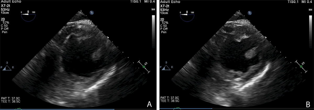
Figure 4
(A) Transgastric transesophageal echocardiography (TEE) short axis view of a failing left ventricle at end systole. (B) TEE short axis view of a failing left ventricle at end diastole. Photo credit Chandrika Garner, MD, Atrium Health Wake Forest University Medical Center
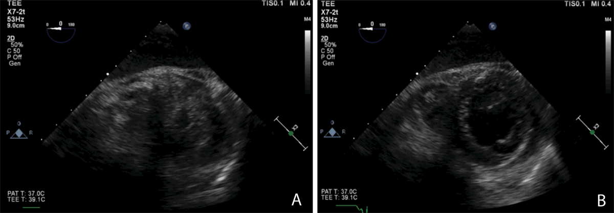
Figure 5
(A) Transgastric short axis transesophageal echocardiography (TEE) view of a hypertrophied, underfilled left ventricle in end systole. (B) Transgastric short axis TEE view of a hypertrophied, underfilled left ventricle in end diastole. Photo credit Chandrika Garner, MD, Atrium Health Wake Forest University Medical Center
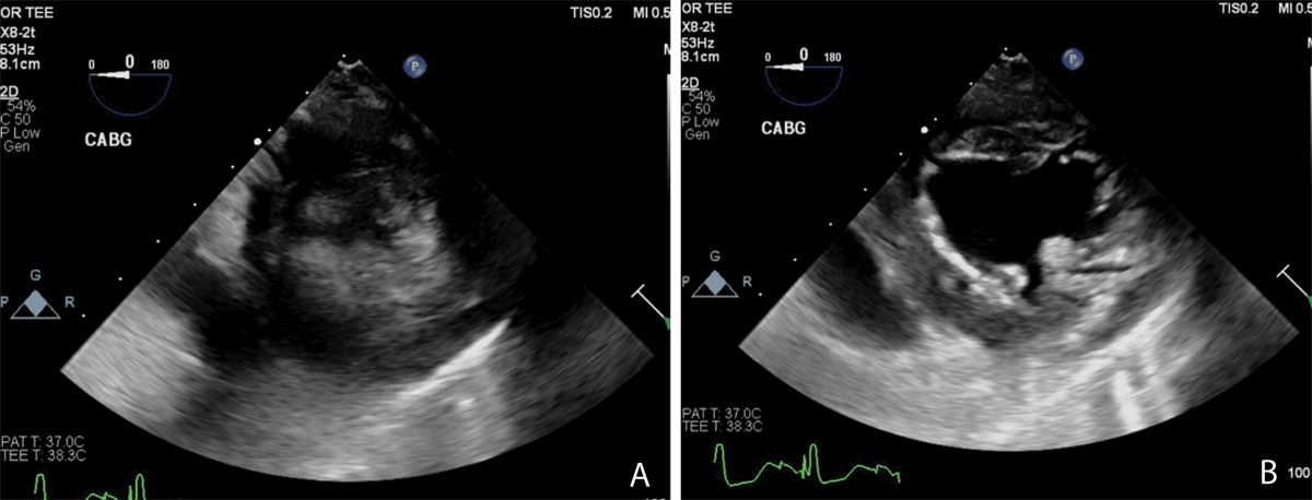
Figure 6
(A) Transgastric short axis transesophageal echocardiography (TEE) view of an underfilled left ventricle with normal function at end systole. This patient was hypotensive and responded to volume administration. (B) Transgastric short axis TEE view of an underfilled left ventricle with normal function at end diastole. Photo credit Kyle Buck, MD, Atrium Health Wake Forest Baptist Medical Center
Using Intraoperative Data to Guide Blood Pressure Management in the Critical Care Unit
Intraoperative hypotension with even brief exposure to a MAP of < 65 mm Hg is known to be associated with an increased risk of organ system failure and mortality.7,42,43,44,45,46 Specifically, intraoperative hypotension is associated with postoperative acute kidney injury (AKI), myocardial injury, stroke, delirium, and mortality.7,45,47,48 Interestingly, this relationship and risk is no different for an absolute MAP < 65 mm Hg or a relative drop from baseline by about 25%.49 Patients who are hypotensive in the operating room are more likely to remain hypotensive in the post-surgical ICU and possibly when they recover on the hospital ward, where it may be undetected for lack of adequate monitoring.43,50,51 An analysis by Khanna et al. showed that a lowest MAP reading less than that compared to a population median MAP of 87 mm Hg in the surgical ICU was significantly associated with the risk of a composite of myocardial injury or mortality. However, intraoperative hypotension had a significant interaction with the primary outcome.52
Even patients with normotension in the operating room may suffer harm associated with trivial new-onset hypotension in the post-surgical ICU.8 Unsurprisingly, in patients undergoing cardiac surgery with CPB, the total duration of intraoperative hypotension per 10-minute exposure of MAP < 65 mm Hg throughout the surgery was statistically significantly associated with the composite primary outcome of stroke, AKI, or death.53 Hypotension pre-CPB can result from decreased venous return, arrythmias due to surgical manipulation, depressed cardiac function, decreased systemic vascular resistance, or induced hypotension for aortic cannulation.54 Despite much emphasis placed on perfusion pressure, there are no guidelines for target perfusion pressure during CPB. According to the 2019 European Adult Cardiac Surgery Guidelines for CPB, it is safe to maintain MAP targets between 50 and 70 mm Hg during CPB.55 However, studies have shown that higher MAP targets during CPB were associated with lower post-bypass lactate levels,56 lower incidence of stroke,13 and decreased risk of AKI.53
It may be prudent to add monitors of cerebral perfusion and oxygenation in patients undergoing cardiac surgery, particularly in those with a history of a cerebrovascular event. A good clinical practice may be to use baseline cerebral oximetry (placed before induction of anesthesia) to monitor trends in the oximeter values with changes in blood pressure and to establish a baseline MAP goal for these patients. Subsequently, this more personalized approach could be used as a targeted postoperative MAP in the ICU to optimize cerebral perfusion pressure.
Transesophageal echocardiography is a good guide to establish volume status intraoperatively and to estimate the patient’s blood pressure goals, cardiac function, and need for pressors versus inotropes.57 Additionally, renal resistive index (RRI) calculated using TEE has shown that an RRI > 0.68 is a good predictor of AKI in patients undergoing cardiac surgery on CPB.58 An important analysis showed significant associations of intraoperative post-CPB RRI elevation with subsequent development of AKI in a cohort of 99 adult cardiac surgery patients.59 However, there remains a lack of evidence to support a higher MAP target in patients with increased baseline RRI, who are presumably at high risk for postoperative AKI and mortality.
Looking to the Future
The latest iteration of blood pressure evaluation pivots from dynamic measurements to prediction and proactive management of hypotension. This is a step beyond our traditional management of hypotension based on reactive responses, which result in time delays of various lengths and may not be adequate to prevent downstream effects.
Machine-learning-derived algorithms that predict hypotension are based on arterial wave form analysis using millions of waveforms from various shock pathophysiological states in model development and external validation.60,61 One such algorithm can predict arterial hypotension with high sensitivity and specificity.61 Davies et al. analyzed this algorithm in 255 patients undergoing major noncardiac surgery and determined a sensitivity and specificity of about 80% 15 minutes preceding hypotension and 85% five minutes prior.62 A recent systematic review by Li et al. evaluated a total of five trials of the hypotension predictive index and determined that it has the potential to reduce frequency and severity of hypotensive episodes when strictly adhering to the protocol.60
Most data surrounding these technologies come from the operating room and support the thought that different varieties of intraoperative hypotension need different treatment, an idea which also could be extrapolated to the ICU within limits.63,64 Randomized trials have reported conflicting evidence for benefits, which may be due to variable adherence to protocol and directions from technology.65,66 Ongoing trials, including the HPI (Hypotension Prediction Index) CARE Trial, are currently underway to examine this technology in cardiac surgery and cardiac critical care.67 Other trials, such as the REACT-SHOCK group in Australia and New Zealand, will examine targeted BP management in the ICU based on baseline (pre-morbid) pressures. Previously, important data suggested that we tend to keep patients with lower pre-morbid pressures on a longer duration of vasopressors, with consequently longer ICU stays, all of which may be unnecessary.68 This and the use of perfusion pressure targets for the future of large interventional pressure targeted trials will be necessary as we grow the evidence base.
Conclusion
We have yet to establish a definitive target for hypotension in different clinical settings. This should prompt us to move away from using a single predefined number for a hypotensive threshold for all patients and towards using a more individualized approach based on patient characteristics. Further, we should consider integrating emerging innovative technologies to assess and monitor an individual patient’s hemodynamic profile, using parameters in addition to blood pressure, before and during admission to a critical care unit. The goal is to optimize tissue perfusion and oxygen delivery rather than targeting a single blood pressure goal as we advance and personalize care for the critically ill.
Key Points
Individualization of mean arterial pressure (MAP) goals is important in the post-cardiac surgery patient, as different comorbidities, operative interventions, and the presence of mechanical circulatory support, amongst other criteria, may influence the MAP required to maintain adequate perfusion.
Almost 10 years after the impressive work by the SEPSISPAM (Sepsis and Mean Arterial Pressure) group, we need to think of trials with appropriate patient populations, targets, and outcomes, as studies have shown that certain vulnerable patient populations may need a higher blood pressure target.
Other blood pressure components, namely mean, systolic, and diastolic pressures, are comparable in their strength of association with organ system injury and intensive care unit (ICU) mortality.
Hospital outcomes, including mortality, are worse in patients with evidence of both hypotension and hypoperfusion.
Analysis comparing perfusion pressure deficits and MAP deficits have been seen to have a nearly similar and strong association with ICU mortality. This is a critical area of focus.
As intensivists, we should move away from a single predefined blood pressure target for all patients and toward a more individualized approach based on patient characteristics.
CME Credit Opportunity
Houston Methodist is accredited by the Accreditation Council for Continuing Medical Education (ACCME) to provide continuing medical education for physicians.
Houston Methodist designates this Journal-based CME activity for a maximum of 1 AMA PRA Category 1 Credit™. Physicians should claim only the credit commensurate with the extent of their participation in the activity.
Click to earn CME credit: learn.houstonmethodist.org/MDCVJ-19.4.
Competing Interests
D. Ashish K. Khanna is on the advisory board for Medtronic, Philips Research North America, and Retia Medical. He serves as a key opinion leader for Edwards Lifesciences, GE Healthcare, Potrero Medical, and Caretaker Medical. He is also funded with a Clinical and Translational Science Institute (CTSI) NIH/NCTAS KL2 TR001421 award for a trial on continuous postoperative hemodynamic and saturation monitoring and from the Hypertension and Vascular Research Center at the Wake Forest School of Medicine. He has previously received grant funding for the Angiotensin II in High Output Shock trial. The other authors have no competing interests to declare.
The department of anesthesiology at Atrium Health Wake Forest is supported by Edwards Lifesciences under a master clinical trials agreement and has received funding from Medtronic, Masimo, Retia Medical, Potrero Medical, and Caretaker Medical.