
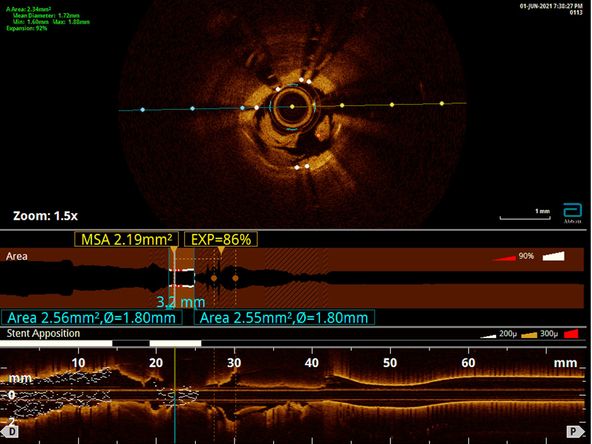
Figure 1
Optical coherence tomography of the right coronary artery showed significant calcification along with severely under-expanded and malapposed stent.
Video 1
Coronary angiography revealed 100% occluded right coronary artery stent causing the patient’s inferior ST-elevation myocardial infarction; see also at https://youtube.com/shorts/ee7qbm-a8G4
Video 2
Final angiographic findings revealed residual 30% to 40% stenosis with Thrombolysis in Myocardial Infarction-3 flow in the right coronary artery; see also at https://youtube.com/shorts/iqwHACzyv4A
Video 3
Coronary angiography revealed 100% occluded right coronary artery stent causing the patient’s recurrent inferior ST-elevation myocardial infarction; see also at https://youtube.com/shorts/ACLrnSVUODc
Video 4
Intravascular ultrasound of the right coronary artery revealed severely calcified lesion with under-expanded and malapposed stent; see also at https://youtube.com/shorts/8oIxN4q0rKo
Video 5
Intravascular ultrasound revealed fragmentation of the calcification and expansion of the stent; see also at https://youtube.com/shorts/HeJhQfgemY4
Video 6
Final angiographic findings revealed residual 0% stenosis with Thrombolysis in Myocardial Infarction-3 flow in the right coronary artery; see also at https://youtube.com/shorts/y1svDHpX4G0
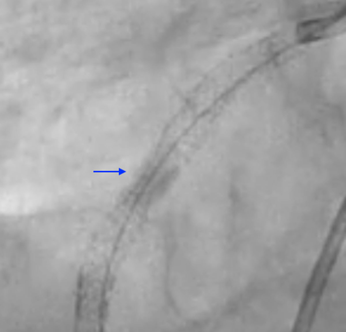
Figure 2
Dry-cine of the right coronary artery stent pre-intravascular lithotripsy.
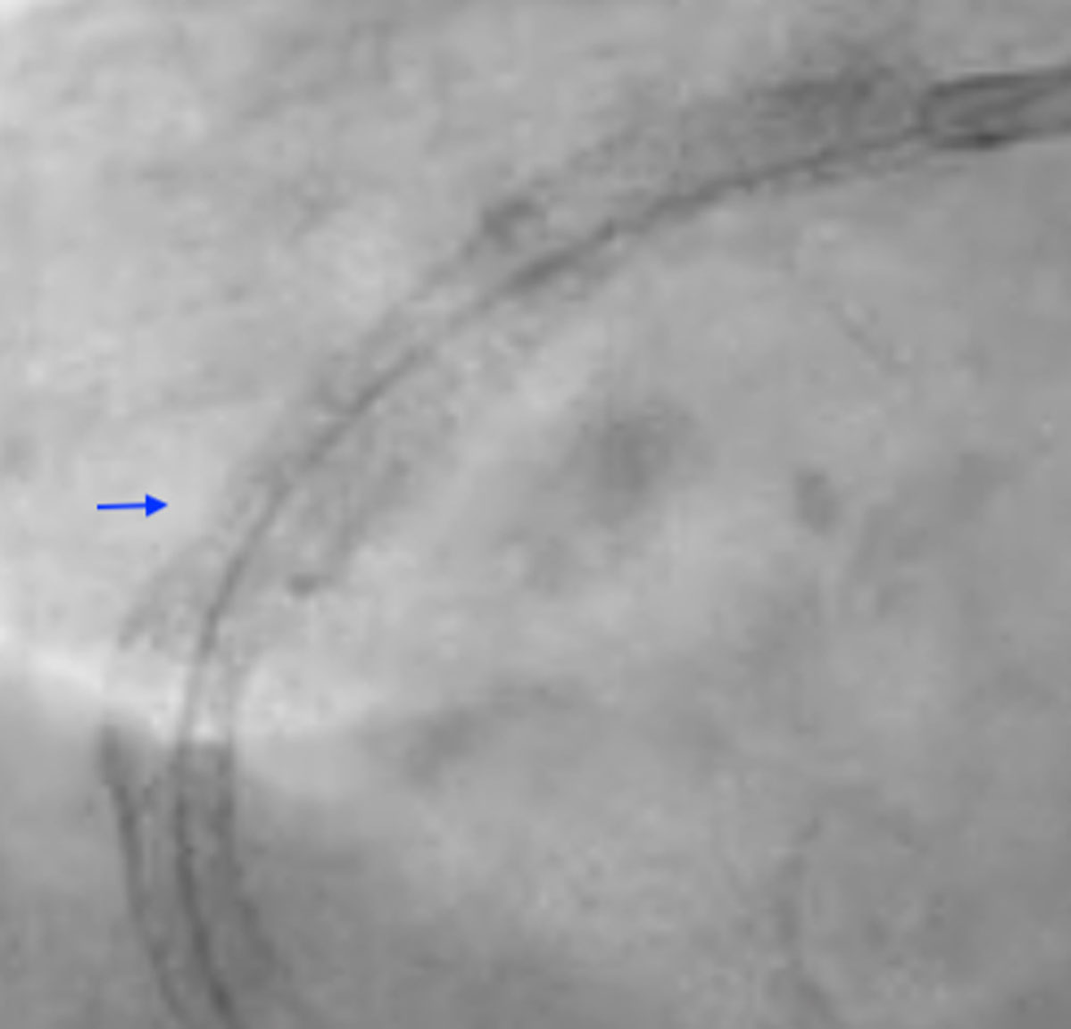
Figure 3
Dry-cine of the right coronary artery stent post-intravascular lithotripsy revealing stent-release.