
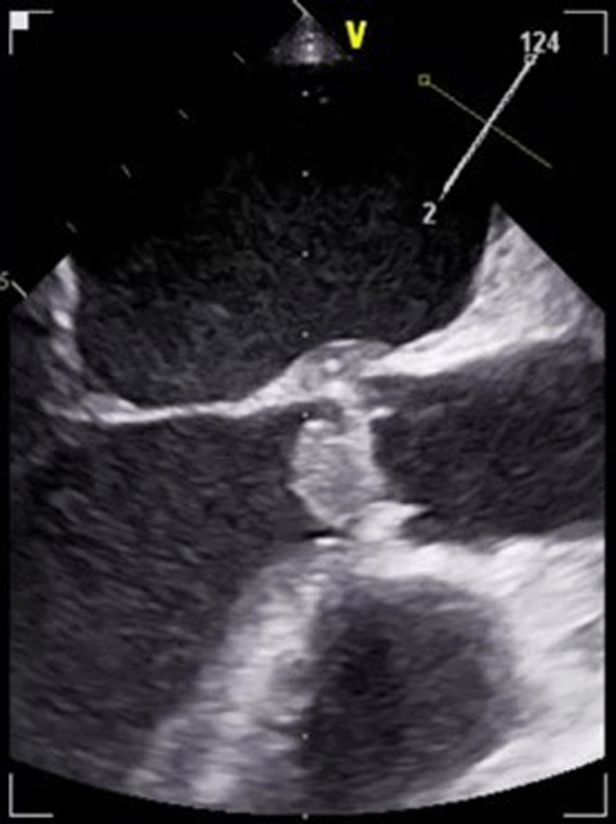
Figure 1
Large echogenic mass can be appreciated on the aortic valve.
Video 1
Transesophageal echocardiography midesophageal aortic valve short axis view (left) and long axis view (right). Large mobile vegetation on the aortic valve with significant left ventricular outflow obstruction; see also at https://youtu.be/nNkrE-YsOKk.
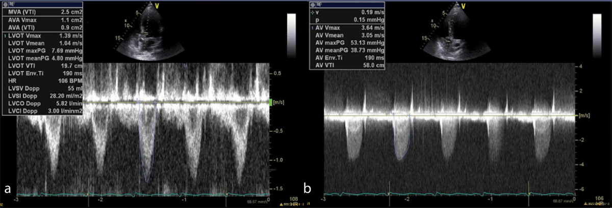
Figure 2
Pulse wave Doppler (2a) and continuous wave Doppler (2b) with evidence of left ventricular outflow obstruction.
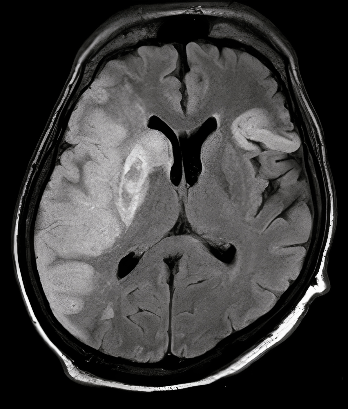
Figure 3
Magnetic resonance imaging of head indicating right-sided middle cerebral artery stroke.