
Figure 1
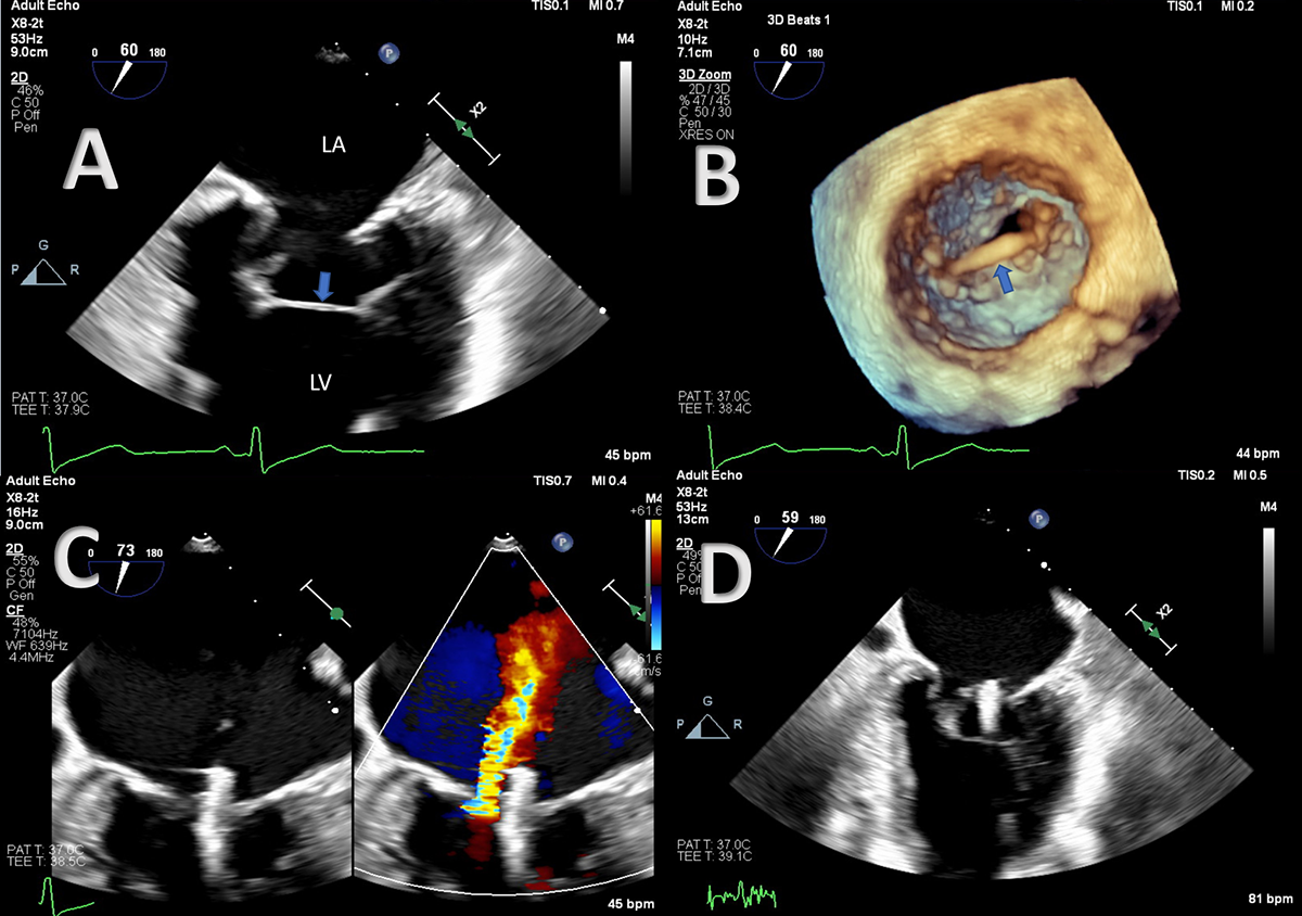
(A) Two-dimensional transesophageal echocardiography (TEE) (mid-esophageal 2-chamber view) shows a transverse chord-like structure (blue arrow) connecting the papillary muscles. (B) Three-dimensional TEE of the mitral valve highlights the transverse tendon (blue arrow). The procedure was successful with (C) residual mild mitral regurgitation, and (D) the tendon remained intact. LA: left atrium; LV: left ventricle.
Video 1
Two-dimensional biplane transesophageal echocardiography, two chamber mid-esophageal view, showing transverse chord-like structure close to mitral valve connecting the papillary muscles; see also at https://youtu.be/rBnvKGOCzgg.
Video 2
Three-dimensional transesophageal echocardiography showing atypical left ventricular false tendon; see also at https://youtu.be/8ScrmhzvQZo.
Video 3
Two-dimensional transesophageal echocardiography showing successful MitraClip implantation with residual mitral regurgitation and the tendon remained intact; see also at https://youtu.be/XPrZcds-wEk.