
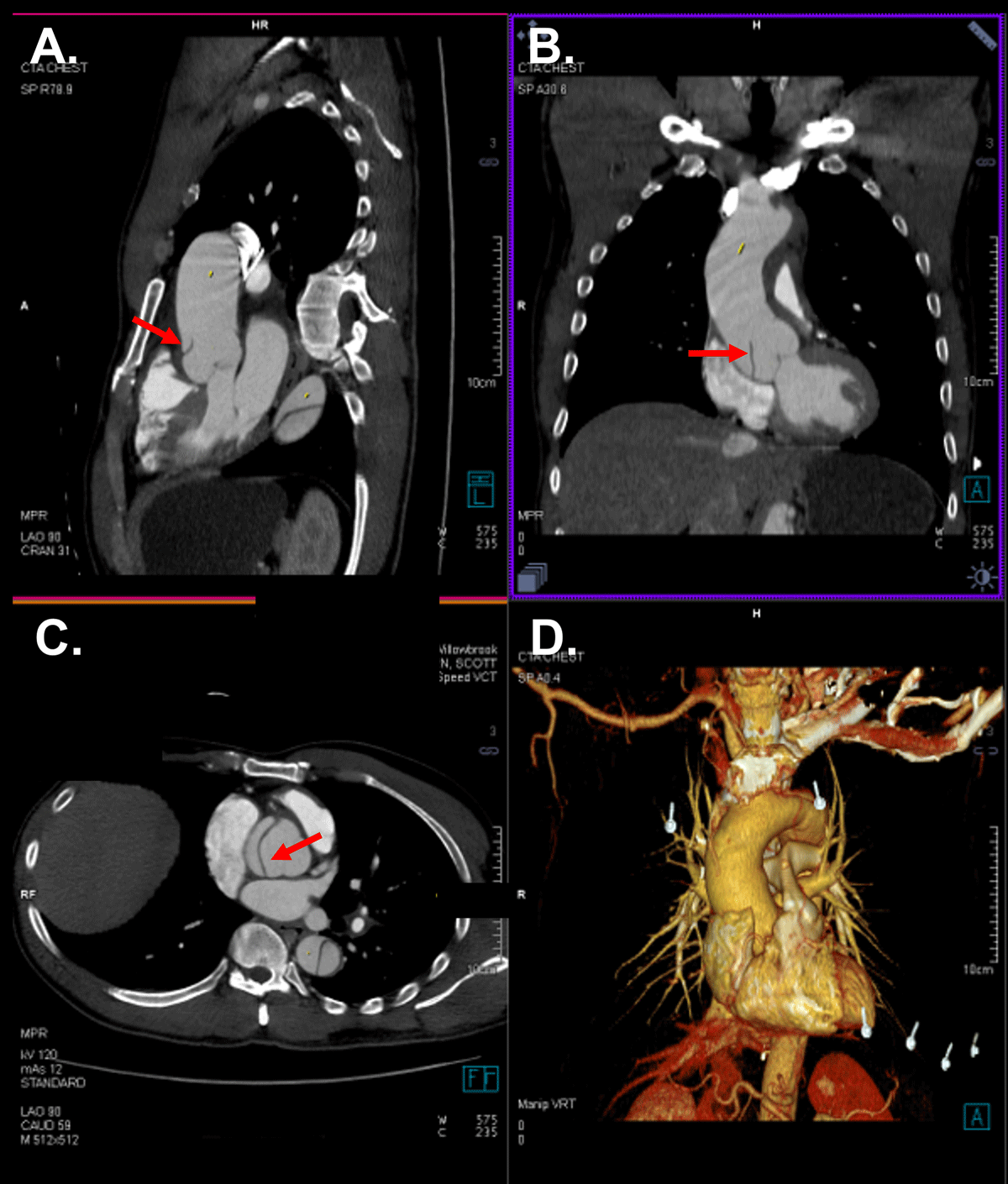
Figure 1
Aortic root dissection. Sagittal (A), coronal (B), and transverse (C) computed tomography showing dissection flap in the aortic root (arrow). Three-dimensional reconstructed computed tomography is provided (D).
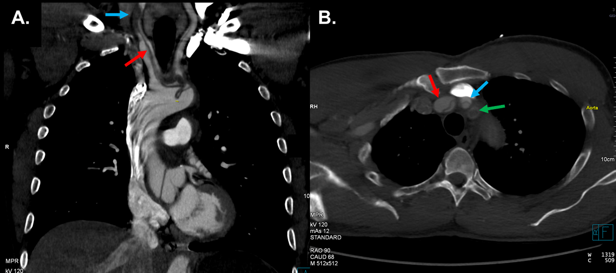
Figure 2
Supra-aortic vessel dissection. Sagittal (A) and transverse (B) computed tomography showing brachiocephalic artery dissection (red arrow) and near complete occlusion of the right common carotid artery (blue arrow) due to thrombosis of the false lumen. Dissection of the right subclavian artery (green arrow) is also seen.
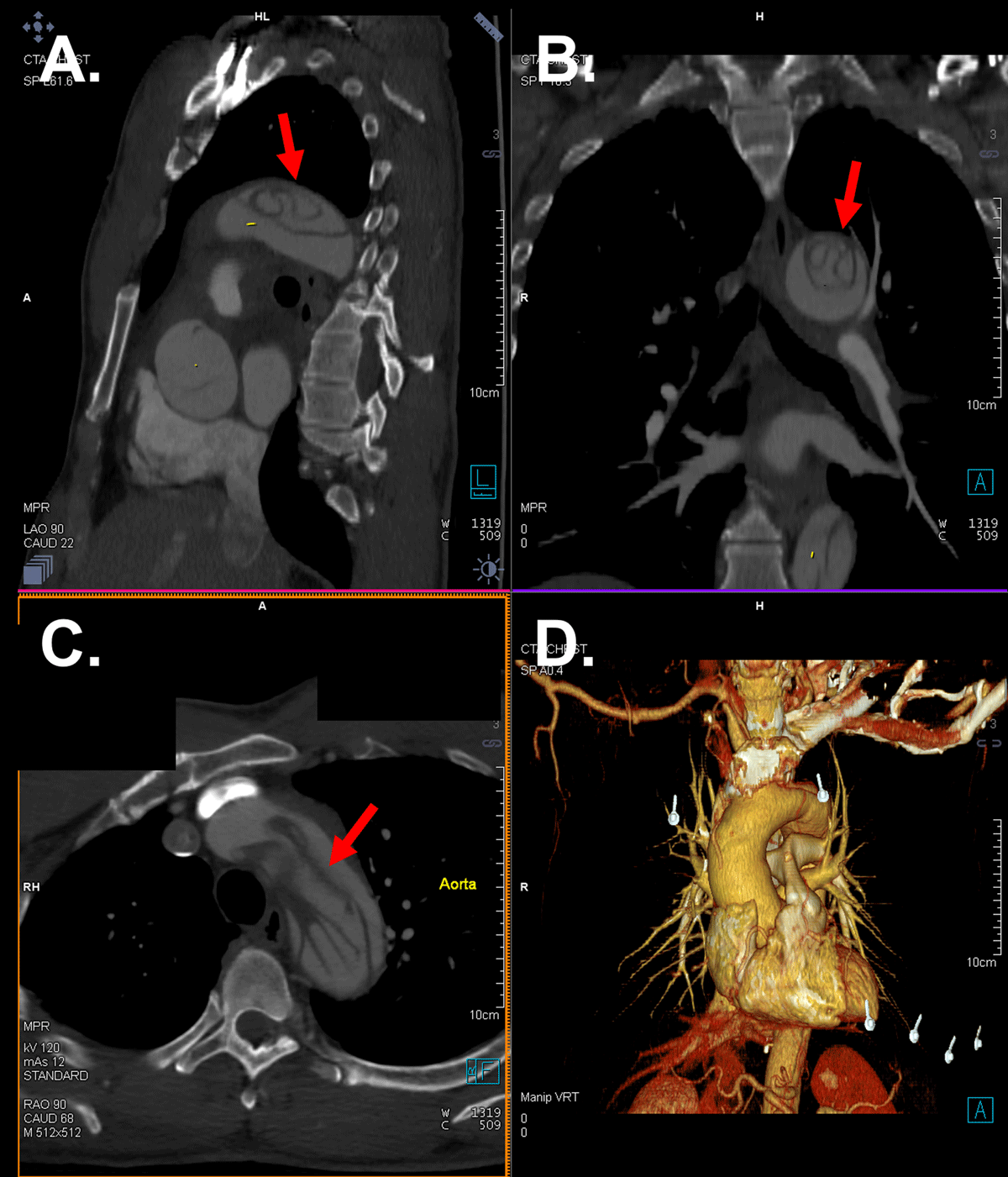
Figure 3
Aortic arch dissection. Sagittal (A), coronal (B), and transverse (C) computed tomography showing complex dissection flap at the level of the aortic arch (arrow). Three-dimensional reconstructed computed tomography is provided (D).
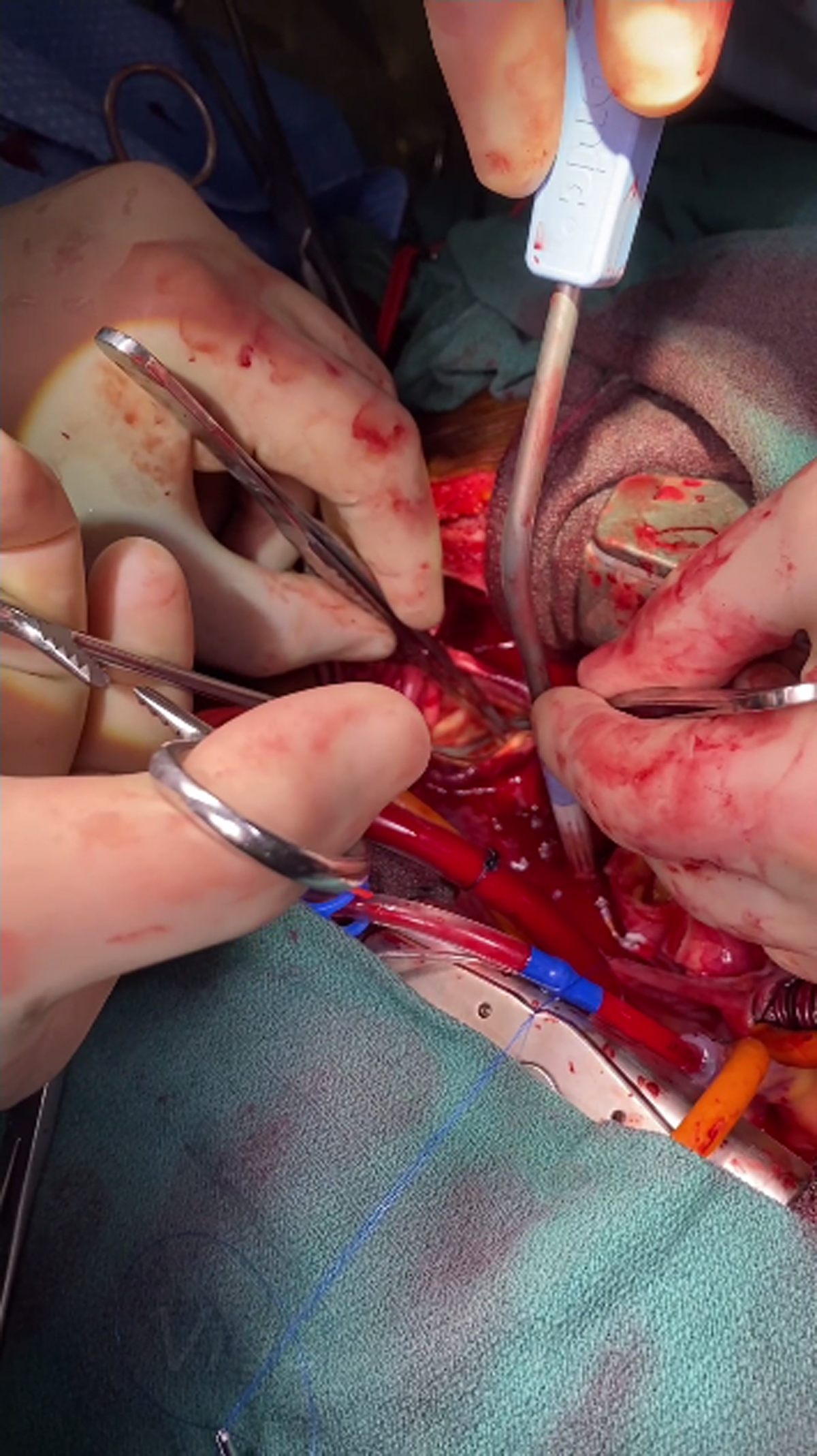
Video 1
Surgical video of the intussuscepted intima being grasped within the distal arch/descending aorta and rolled back on itself for realignment; see also at https://youtube.com/shorts/f4mnD3ucmgY.