
Table 1
Quantitative and qualitative criteria for grading of mitral regurgitation severity. EROA: effective regurgitant orifice; LVOT: left ventricular outflow tract; PHT: pressure half time
| CRITERIA | MILD (1+) | MODERATE (2+) | MODERATE-SEVERE (3+) | SEVERE (4+) |
|---|---|---|---|---|
| QUANTITATIVE | ||||
| Regurgitant volume (mL) | < 30 | 30-40 | 45-59 | ≥ 60 |
| Regurgitant fraction (%) | < 30 | 30-39 | 40-49 | ≥ 50 |
| EROA (cm2) | < 0.1 | 0.1-0.2 | 0.2-0.29 | ≥ 0.3 |
| QUALITATIVE | ||||
| Jet width in LVOT | Small in central jets | Intermediate | Large in central jets | |
| Flow convergence | None or very small | Intermediate | Large | |
| Jet density | Incomplete or faint | Dense | Dense | |
| Jet deceleration rate (PHT, msec) | Faint slow, > 500 | Medium, 500-200 | Steep, < 200 | |
| Diastolic flow reversal | Brief, early diastolic | Intermediate | Prominent holodiastolic | |
Table 2
Major inclusion and exclusion echocardiographic criteria for transcatheter edge-to-edge repair derived from the COAPT and EVEREST II trials.
| MAJOR INCLUSION CRITERIA |
|---|
| The primary regurgitant jet originates from malcoaptiation of the A2 and P2 scallops of the mitral valve (MV) |
| MAJOR EXCLUSION CRITERIA |
| MV orifice area < 4.0 cm2 |
| Width of the flail segment ≥ 15 mm or flail gap ≥ 10 mm |
| Coaptation depth > 11 mm or vertical coaptation length is < 2 mm |
| Severe mitral annular calcification |
| Evidence of calcification in the grasping area of the A2 and/or P2 scallops |
| Presence of a significant cleft of A2 or P2 scallops |
| Prior MV surgery or valvuloplasty |
| Echocardiographic evidence of intracardiac mass, thrombus, or vegetation |
| History of or active endocarditis or rheumatic heart diseases |
| History of atrial septal defect or patent foramen ovale associated with clinical symptoms |
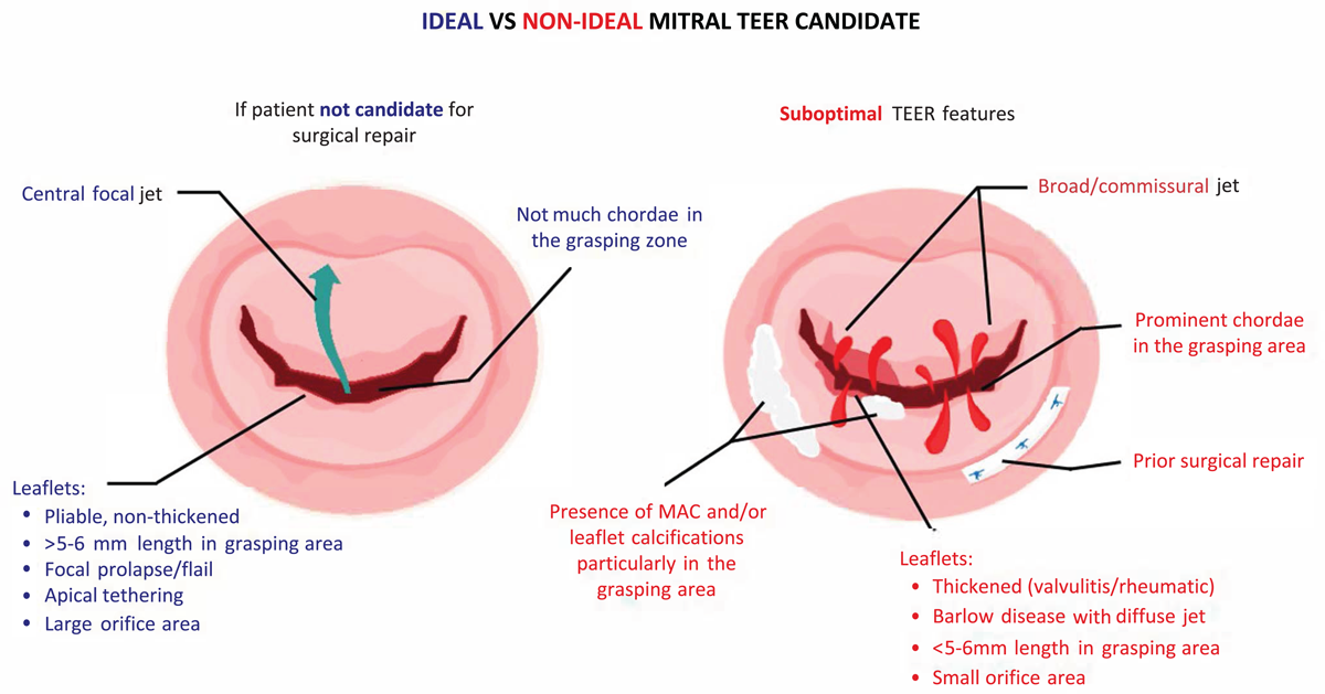
Figure 1
Summary of ideal versus nonideal candidates for transcatheter edge-to-edge repair.
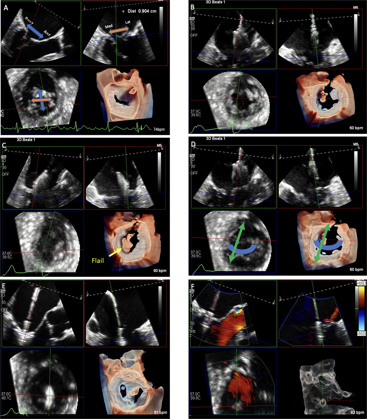
Figure 2
Multiplanar reconstruction (MPR) of the mitral valve using transesophageal echocardiography for assessment and procedural guidance. Multiplanar assessment of the mitral valve (A) is done by visualizing the long-axis view (top left plane) of the mitral valve on which anterior and posterior locations could be localized, the bi-commissural (top right) plane where medial and lateral coordinates can be determined; both of these planes will determine the small axis plane (bottom left) and its 3D reconstruction (bottom right). During the procedure, real-time MPR allows (B) visualization and adjustment of the device trajectory, (C) positioning of the device according to disease location, (D) orientation of device for optimal grasping, (E) advancement of the device and grasping, and (F) evaluation of device attachment and residual disease.
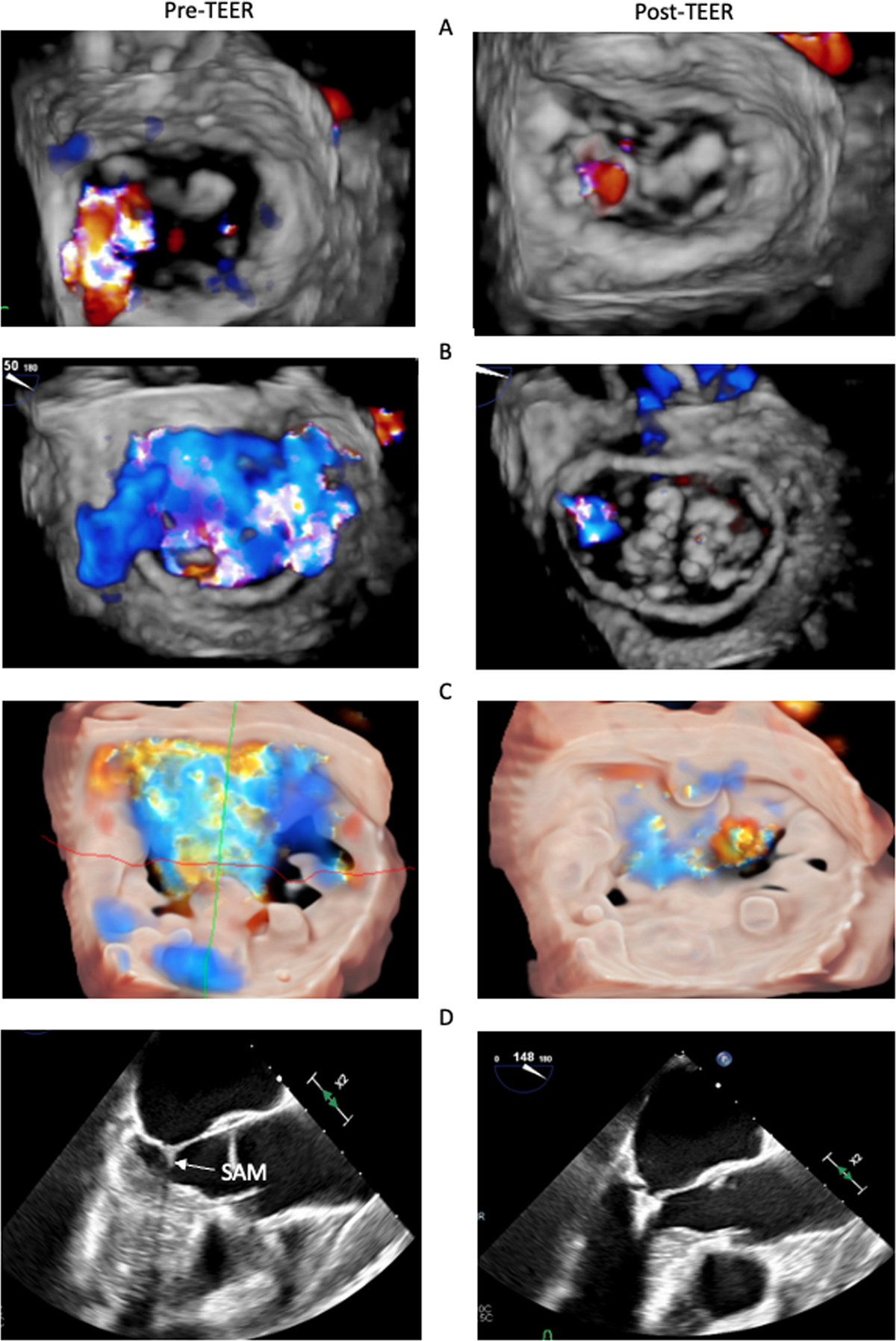
Figure 3
Pre- and post-transcatheter edge-to-edge repair (TEER) echocardiography in challenging cases of TEER in (A) commissural mitral regurgitation; (B) prior ring annuloplasty; (C) severe mitral annular calcification; and (D) hypertrophic cardiomyopathy with left ventricular outflow tract obstruction due to systolic anterior motion (SAM).
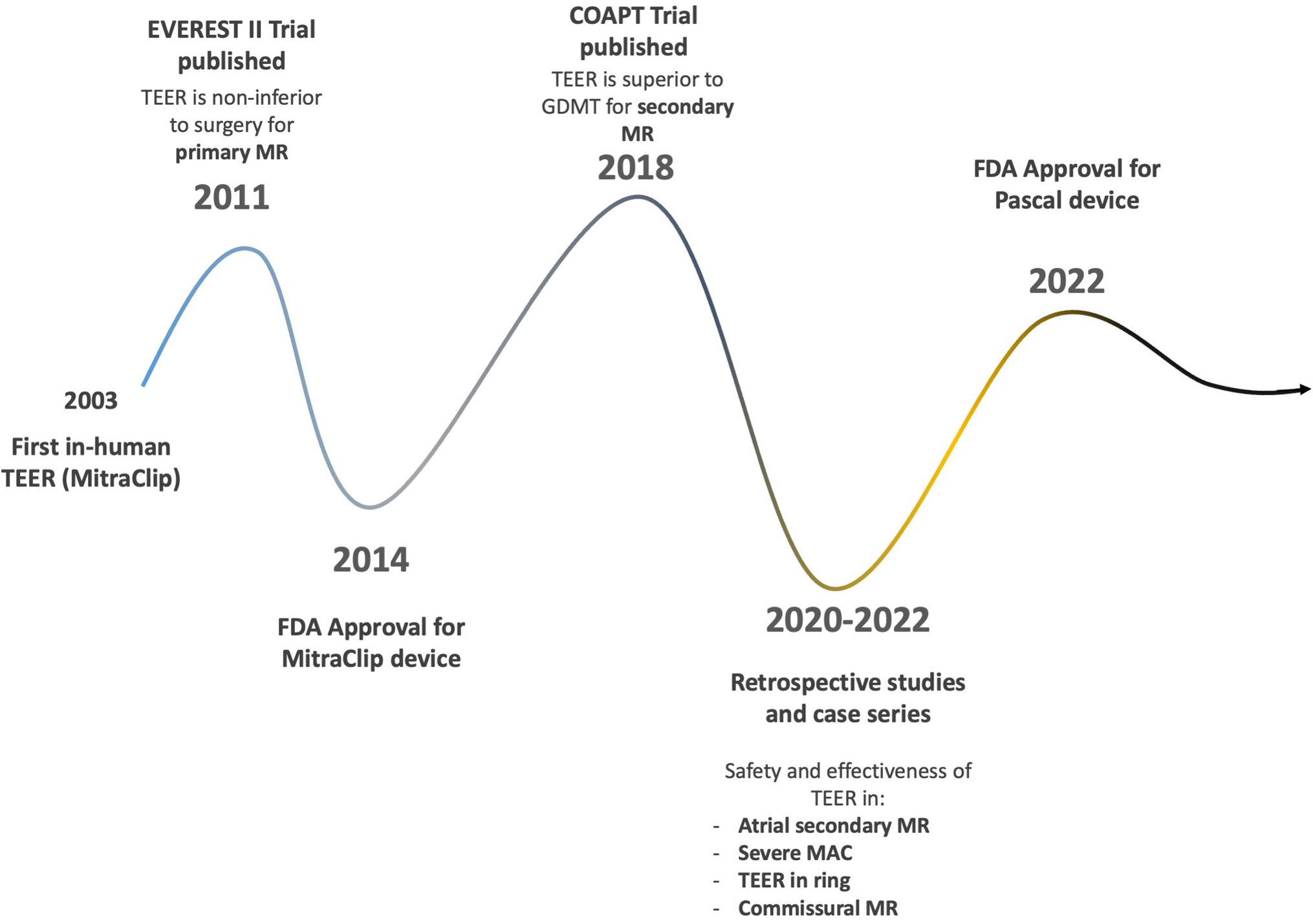
Figure 4
Timeline of scientific and technological advancements of transcatheter edge-to-edge repair since the first in-human procedure. MR: mitral regurgitation; GDMT: goal-directed medical therapy; TEER: transcatheter edge-to-edge repair; MAC: mitral annular calcification