
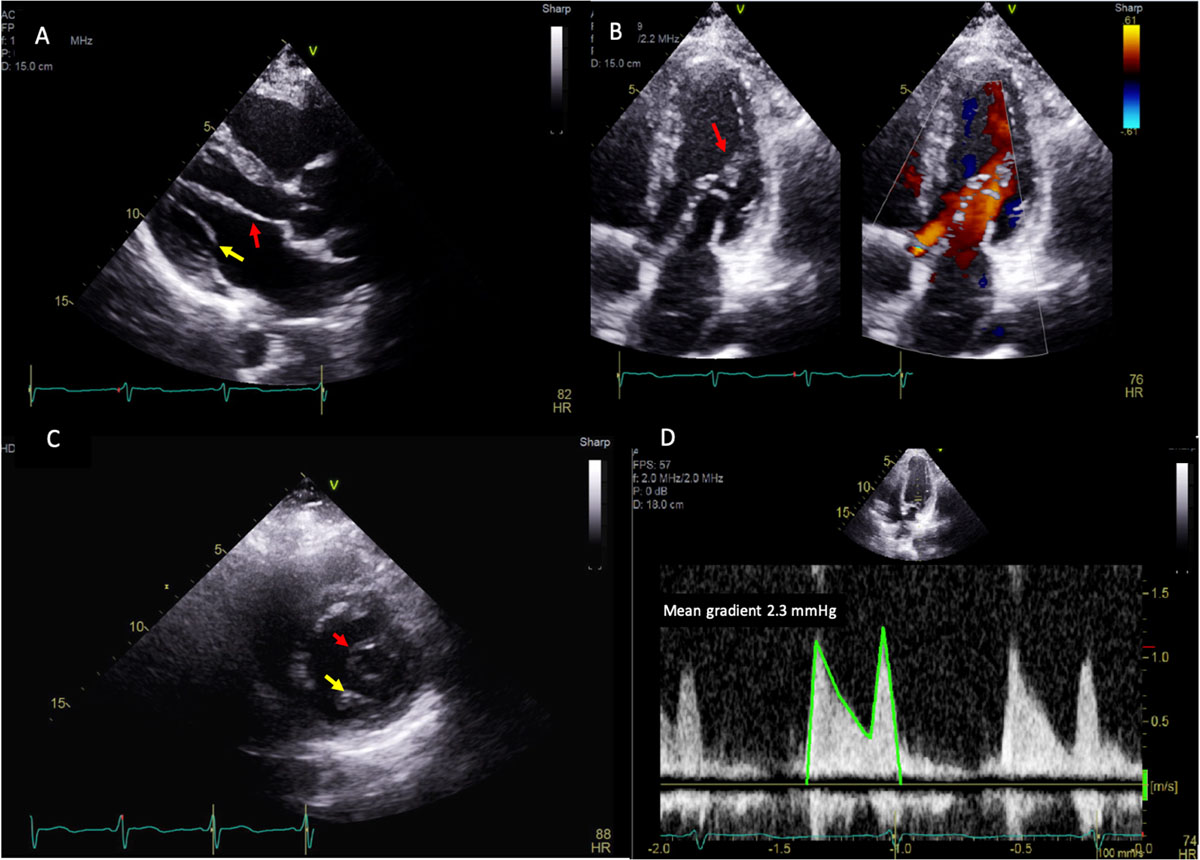
Figure 1
(A) Parasternal long view on transthoracic echo showing thickened anterior (red arrow) and posterior mitral valve (yellow arrow) leaflets. (B) Apical long-axis view showing pear-shaped eccentric opening of the mitral valve with short chordae tendinea attached to the anterolateral papillary muscle (red arrow) and transmitral flow during diastole. (C) Parasternal short-axis view showing an eccentric mitral valve orifice (red arrow) predominately oriented towards the anterolateral papillary muscle and a remnant of the posteromedial papillary muscle (yellow arrow). (D) Spectral Doppler showing mean gradient of 2.3 mm Hg at a heart rate of 74 beats/min across the mitral valve in diastole.