
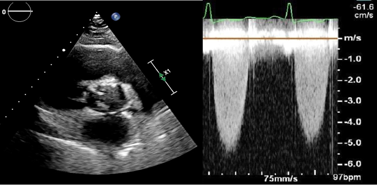
Figure 1
Echocardiogram of short axis view showing a bicuspid valve with increased velocity across the valve; see also at https://youtu.be/4C2kncWIOqM.
Video 1
Echocardiogram of short axis view showing a bicuspid valve with increased velocity across the valve; see also at https://youtu.be/4C2kncWIOqM.
Video 2
Echocardiogram of long axis color Doppler view showing a bicuspid valve with severe stenosis; see also at https://youtu.be/Y4cAeFUFoPs.
Video 3
Coronary angiography of fluoroscopy showing a coronary artery fistula arising from the proximal right coronary artery; see also at https://youtube.com/shorts/yZ3eEaNSuPw.
Video 4
A coronary angiography of fluoroscopy showing a coronary artery fistula arising from the mid-left anterior descending artery and draining into the main pulmonary artery; see also at https://youtube.com/shorts/gHFhO8Gxb8c.
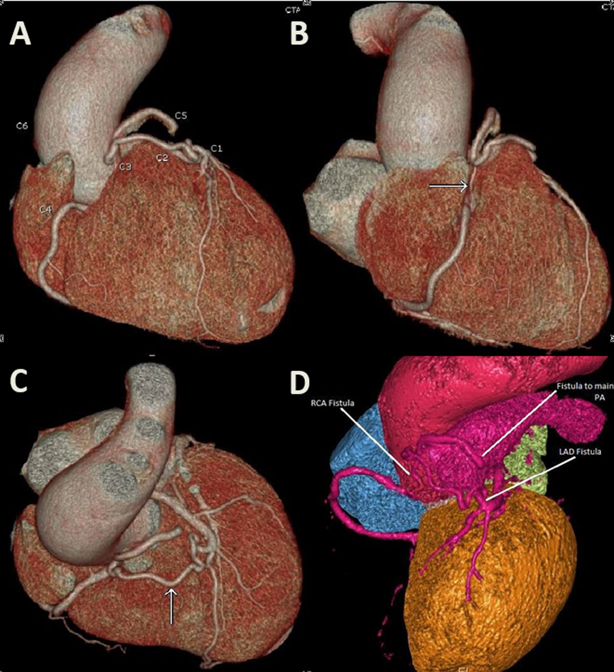
Figure 2
A coronary computed tomography angiography 3-dimensional (3D) reconstruction showing a coronary artery fistula arising from the left anterior descending artery joining another fistula arising from the right coronary artery, both draining into the main pulmonary artery. (A) C1: left anterior descending artery; C2: left anterior descending artery fistula; C3: right coronary artery fistula; C4: right coronary artery; C5: drainage into the pulmonary artery; C6: ectasia of the ascending aorta; (B) arrow: fistula connecting to the right coronary artery; (C) arrow: fistula connecting to the left anterior descending artery; (D) 3D reconstruction of the fistula excluding the right ventricle.
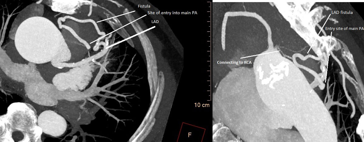
Figure 3
A coronary computed tomography angiography axial view showing the course of the fistula.