
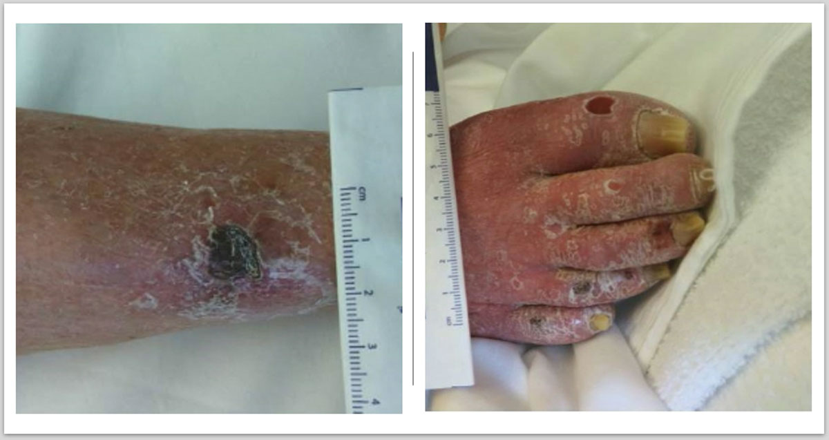
Figure 1
Preoperative image of right lower extremity showing necrotic ulcers in the leg and toes.
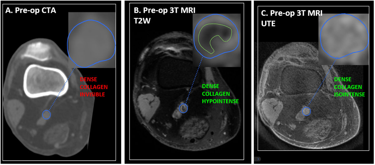
Figure 2
Preoperative (pre-op) imaging with computerized tomographic angiography (CTA) and magnetic resonance imaging (MRI). CTA shows an occluded popliteal artery isodense to the reference muscle tissues (A). MRI shows a lesion that is hypointense in T2-weighted (B)and isointense in ultrashort echo time (C), characteristic of dense collagen.
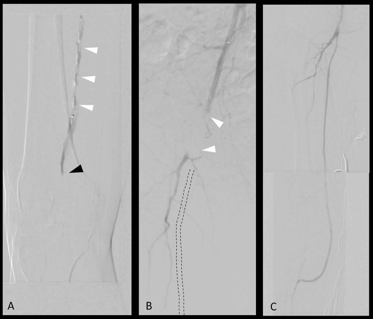
Figure 3
Intraoperative angiograms. (A) Failed attempt to recanalize occlusion (black arrow) with subintimal dissection (white arrows). (B) Flow-limiting dissection of the femoral artery due to multiple recanalization attempts. (C) Successful restoration of flow after femoropopliteal bypass.
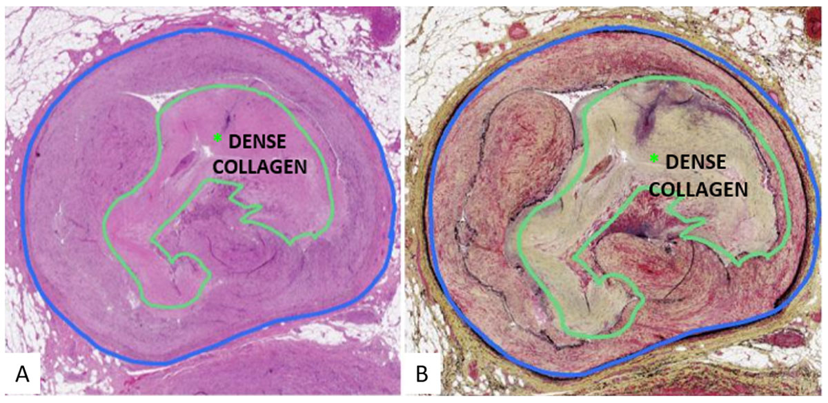
Figure 4
Histology of popliteal artery lesion showing dense collagen composition of atheromatous plaque.