
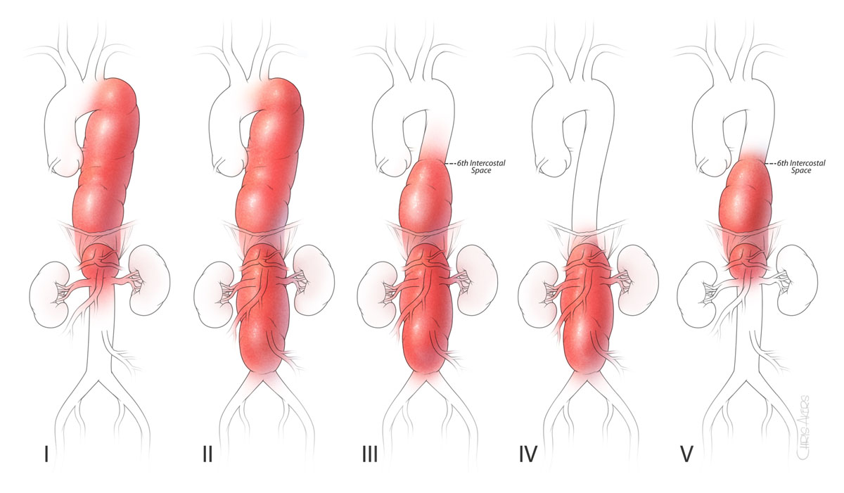
Figure 1
Classifications of thoracoabdominal aortic aneurysms—the scheme of thoracoabdominal aortic aneurysms depicted by extent of aneurysm.
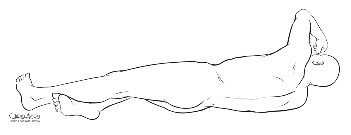
Figure 2
Positioning and skin incision. Note the scapulars are at 90 degrees to the table but hips are rotated for abdominal and femoral exposures.
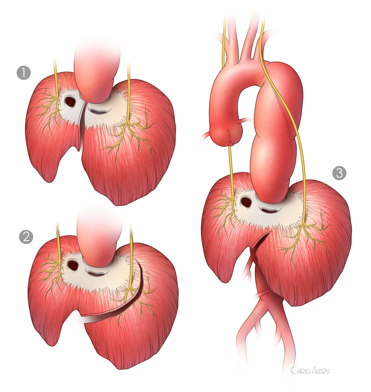
Figure 3
Diaphragm splitting. (1) Radial division, (2) lateral division, and (3) partial lateral division. Partial lateral division of the diaphragm with sparing of the phrenic nerve has been associated with enhanced postoperative recovery of diaphragmatic and pulmonary function. Note that we cut only one-third of the circumference.
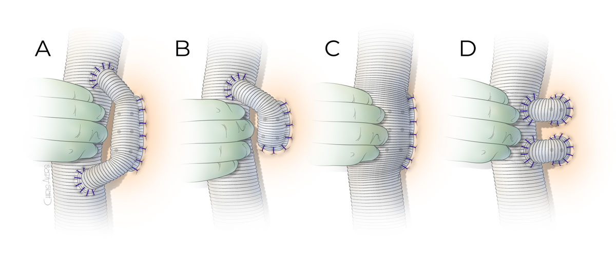
Figure 4
Intercostal artery reattachment techniques. (A) A 14-mm Dacron graft is anastomosed to the anterolateral surface of the main body graft as a loop to reconstruct the intercostal arteries in side-to-end fashion. (B) Similar to the loop graft but the proximal end may not be anastomosed back to the main body graft if reconstructing less than three pairs of intercostal arteries. The anastomosis to the intercostal arteries is performed in end-to-end fashion by beveling the 14-mm graft. (C) A side hole is made large enough to accommodate the intercostal arteries to be reattached and anastomosed as an island patch in side-to-side fashion. (D) A 12-mm ×14-mm Dacron graft is used to reattach each pair of intercostal arteries in an end-to-end fashion.
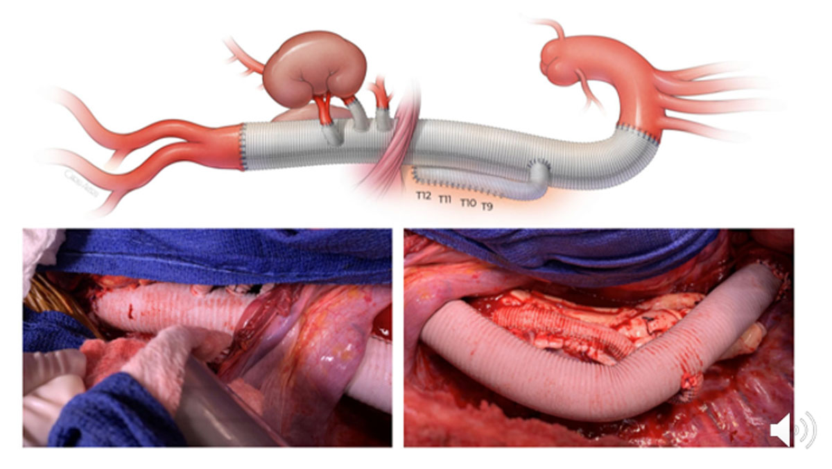
Figure 5
Extent II thoracoabdominal aortic aneurysm repair with side-branched thoracoabdominal aortic graft (STAG).
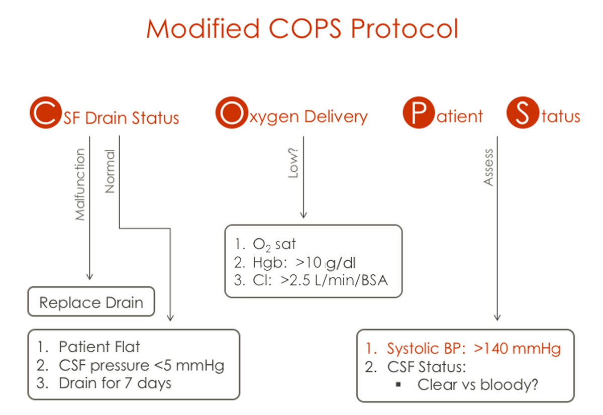
Figure 6
When delayed paraplegia occurs, use a modified COPS protocol: Limitless Cerebrospinal fluid drainage to maintain pressure below 5 mm Hg; Oxygen delivery is optimized by keeping hemoglobin above 10 g/dL and cardiac index above 2.5 mL/min/body surface area m2; and optimize Patient Status by elevating the systolic blood pressure above 140 mm Hg.