
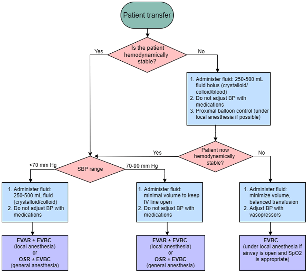
Figure 1
Algorithm for preoperative blood pressure and volume management. Hemodynamic stability is defined as patient consciousness with no fluid/pressors to maintain blood pressure between 70-90 mm Hg for at least 5 minutes. BP: blood pressure; EVAR: endovascular aortic repair; EVBC: endovascular balloon control; OSR: open surgical repair; SBP: systolic blood pressure.
Table 1
Important computerized tomography angiogram findings prior to treatment of RAAA.2,4 EVAR: endovascular aortic repair; OSR: open surgical repair; AAA: abdominal aortic aneurysm; RAAA: ruptured abdominal aortic aneurysm
| EVAR | OSR | OTHER CONSIDERATIONS REGARDLESS OF EVAR OR OSR |
|---|---|---|
| Diameter (< 32 mm), angle (< 60°) and length (≥ 10 mm depending on type of endograft) of aneurysmal neck | Extent of healthy artery free of calcifications and aneurysms | Superior mesenteric artery: level of aortic origin, presence of stenosis/obstruction |
| Diameter of aortic bifurcation (≥ 17 mm) | Distal disease | |
| Diameter of common iliac arteries (≥ 5 mm) | Free vs contained RAAA | |
| Diameter of external iliac arteries (≥ 5 mm) | Presence of retro-aortic left renal vein | |
| Diameter of femoral arteries without excessive tortuosity or calcifications (access point) | Thoracoabdominal aneurysm, inflammatory AAA, horseshoe kidneys |
Table 2
Postoperative complications of ruptured abdominal aortic aneurysm repair and treatment recommendations.4,12,13,14,15,16,17,18,19,20,21,22,23,24 BNP: brain natriuretic peptide; BUN: blood urea nitrogen; CBC: complete blood count; CT: computed tomography; CSF: cerebrospinal fluid; CXR: chest X-ray; ECG: electrocardiogram; ESRD: end-stage renal disease; FiO2: fraction of inspired oxygen; HLA: human leukocyte antigen; IAP: intra-abdominal pressure; INR: international normalized ratio; IMA: inferior mesenteric artery; IVC: inferior vena cava; LMWH: low-molecular weight heparin; MAP: mean arterial pressure; PaO2: arterial partial pressure of oxygen; PEEP: positive end-expiratory pressure; PCR: polymerase chain reaction; PT: prothrombin time; PTT: partial thromboplastin time; SMA: superior mesenteric artery; SSEP: somatosensory evoked potentials; SQ: subcutaneous; tcMEP: transcranial motor evoked potentials; TEG: thromboelastography; THROMBINS2: thienopyridines, renin-angiotensin system blockade, oxygen, morphine, beta-blockers, invasive cardiac interventions, nitroglycerin, statin/salicylate; VV ECMO: veno-venous extracorporeal membrane oxygenation
| ORGAN/SYSTEM | COMPLICATION | PRESENTATION/MONITOR | PREVENTION | TREATMENT |
|---|---|---|---|---|
| Central nervous system | Spinal cord ischemia4,12 | Neurological symptoms, spinal cord (neurophysiologic) monitoring: SSEP and tcMEP, CSF drain monitoring, BP, oxygenation | Avoid sustained hypotension: intraoperative MAP > 80 mm Hg | Increase MAP using IV fluid bolus up to 2 L, vasopressors, inotropes. Spinal drain, increase hemoglobin, oxygenation. |
| Cardiovascular | Cardiac dysfunction4,14,15 | ECG, echocardiograms, serum troponin, serum BNP, lactate | Avoid volume overload (give furosemide post-op day 3 if necessary). Avoid tachycardia. Intraoperative communication with anesthesia during reperfusion to avoid excessive ischemic reperfusion injury. | Practice THROMBINS2 |
| Bleeding | CBC (hemoglobin, platelet), fibrinogen, INR/PT/PTT, calcium, TEG, BUN | Correct coagulopathy early. Address surgical bleed early. Appropriate surgical technique (avoid venous injury) prior to heparinization. Avoid hemodilution. Avoid excessive/prolonged heparinization. | If surgical bleeding, need early mobilization to OR prior to the patient becoming coagulopathic. With bleeding patient, practice balanced transfusion. For ESRD patients, give desmopressin. | |
| Deep venous thrombosis4,16,17 | Lower/upper extremity swelling, D-dimer, venous ultrasound | Early mobilization, intermittent pneumatic compression, subcutaneous heparin/LMWH, removal of central access catheters, when possible. DVT prophylaxis (after successful repair and stable hemoglobin for 24 h). | Heparin/LMWH (SQ), Fondaparinux (SQ), warfarin. If unable to anticoagulate, place IVC filter. | |
| Access complication | Limb ischemia due to access site/limb thrombosis, groin complications (seroma, hematoma, infection), which can be associated with drainage, erythema, wound dehiscence | Hourly neurovascular assessment for signs of ischemia, daily assessment for wound complication | Recognize the access site complication and address it in a timely manner by returning to the OR. | |
| Pulmonary | Transfusion-associated cardiogenic overload18 | Elevated serum BNP, acute/worsening pulmonary edema, cyanosis, hypoxia with absence of other specific causes, unexplained cardiovascular changes | Slower infusion rate, preemptive diuretics | Diuretics |
| Transfusion-related acute lung injury18 | Increased oxygen demand, pink/frothy secretions from endotracheal tube, fever, hypotension, cyanosis | Donor screening/deferral for anti-HLA antibodies | Supportive treatment | |
| Ventilator-associated pneumonia 4,21,22 | Development within 48 hours of endotracheal intubation. Respiratory decline, lung infiltrates (CXR), fever, cough. Blood cultures, sputum cultures, PCR testing from nasal swabs. | Elevation of head of the bed 30-45° to prevent aspiration. Oral chlorhexidine. Oral care, hospital hygiene measures (handwashing), timely vaccinations for patients and providers. Reduce number of days on mechanical ventilation. | Empiric antibiotic therapy based on institution-specific antibiogram. | |
| Acute respiratory distress Syndrome4,18,19,20,23,24 | Lung injury onset within 1 week of clinical insult, with symptoms unexplained by cardiac failure or fluid overload. Bilateral, diffuse opacities (CXR/CT). Respiratory failure. Severe hypoxemia. Decreased PaO2/FiO2 ratio (≤ 300 mm Hg). | On mechanical ventilation, maintain low tidal volume (≤ 6 mL/kg predicted body weight). Limit blood transfusion. Reduce number of days on mechanical ventilation. | Noninvasive positive pressure ventilation, low plateau pressures (< 30 cm H2O), permissive hypercapnia, appropriate titration of PEEP (≥ 5 cm H2O), prone ventilation. Continuous IV infusion of neuromuscular blockers. Discuss VV ECMO. | |
| Gastrointestinal | Abdominal compartment syndrome4 | Difficulty ventilating patient, sustained IAP ≥ 20 mm Hg while patient is paralyzed. | Preemptive diuretics; assess for hemorrhage. | Decompression laparotomy. Diuretics, dialysis/hemofiltration, intravenous paralytic agents. |
| Ischemic colitis4 | Abdominal distension and/or pain, fevers, and early bowel movements (post-op day 0/1). Leukocytosis. Persistent acidosis. Diagnose with emergent Flex Sig. | Avoid strong vasoconstrictors (ie, vasopressin or phenylephrine). Revascularize IMA or diseased SMA. | Early recognition by early Flex Sig. Exploratory laparotomy with possible bowel resection. Supportive therapy (fluids, BP support, bowel rest, antibiotics, NG tube). | |
| Renal | Acute kidney injury4 | FENa > 1%, muddy brown casts in urine sediment, low urine output, anasarca. | Minimize vasopressors, avoid prolonged suprarenal aortic cross-clamping, avoid nephrotoxic drugs | Renal replacement therapies (ie, dialysis) |
| Musculoskeletal | Lower extremity ischemia | Physical exam and noninvasive vascular studies. | Intraoperative heparin | Therapeutic anticoagulation, thrombectomy, fasciotomies for extremity compartment syndrome. |
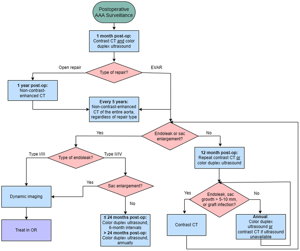
Figure 2
Algorithm for postoperative ruptured aortic abdominal aneurysm surveillance based on Society of Vascular Surgery guidelines and Houston Methodist Hospital protocol.2 CT: computed tomography; EVAR: endovascular aortic repair; OR: operating room.