
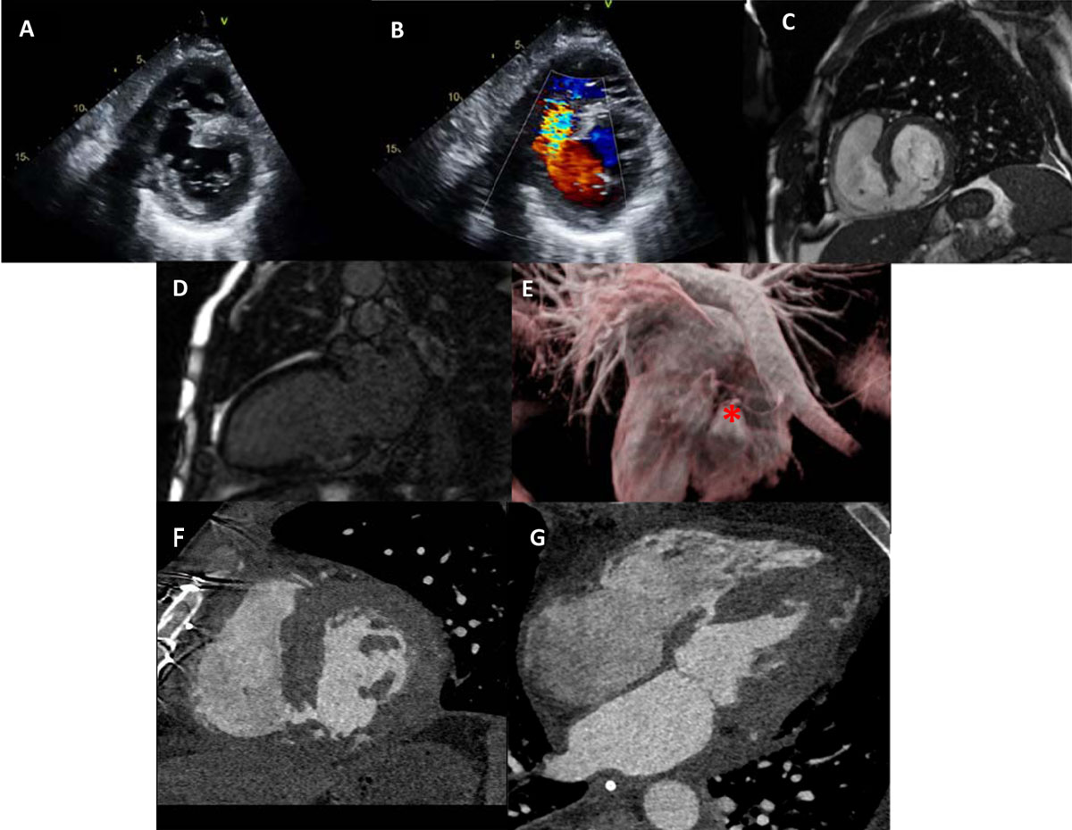
Figure 1
(A) Transthoracic echocardiogram of basal short-axis view showing inferoseptal ventricular septal rupture (VSR) and (B) color Doppler of inferoseptal VSR with left-to-right shunt. (C) Cardiac magnetic resonance imaging (CMR) of basal short-axis view showing inferoseptal VSR and (D) 2-chamber view of nonviable basal inferior wall. (E) Cardiac magnetic resonance of 3-dimensional reconstruction showing inferior aspect of the heart with inferior wall aneurysm (red asterisk). (F) Computed tomography of residual ventricular septal defect showing short-axis view and (G) 4-chamber view.
Video 1
Transthoracic echocardiogram, 2D apical four-chamber view, short-axis shows ventricular septal rupture, see also at https://youtu.be/Lqozswl-tF0.
Video 2
Transthoracic echocardiogram, color Doppler apical four-chamber view, shows ventricular septal rupture left-to-right shunt, see also at https://youtu.be/uMbyqaZt70I.
Video 3
Cardiac magnetic resonance imaging, cine short-axis view, shows ventricular septal rupture, see also at https://youtube.com/shorts/sCPeIJL-TCw.
Video 4
Cardiac magnetic resonance imaging, velocity encoding short-axis view, shows ventricular septal rupture defect, see also at https://youtube.com/shorts/iD4u8PXWMyk.