
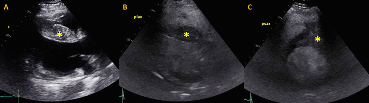
Figure 1
Preoperative transthoracic echocardiogram. (A) Transthoracic echocardiogram without contrast and (B, C) with intravenous contrast-enhancing agent. (B) Parasternal long axis view demonstrating asymmetric septal thickening/mass with contrast enhancement and (C) protrusion of the mass into the right ventricular outflow tract on parasternal short axis.
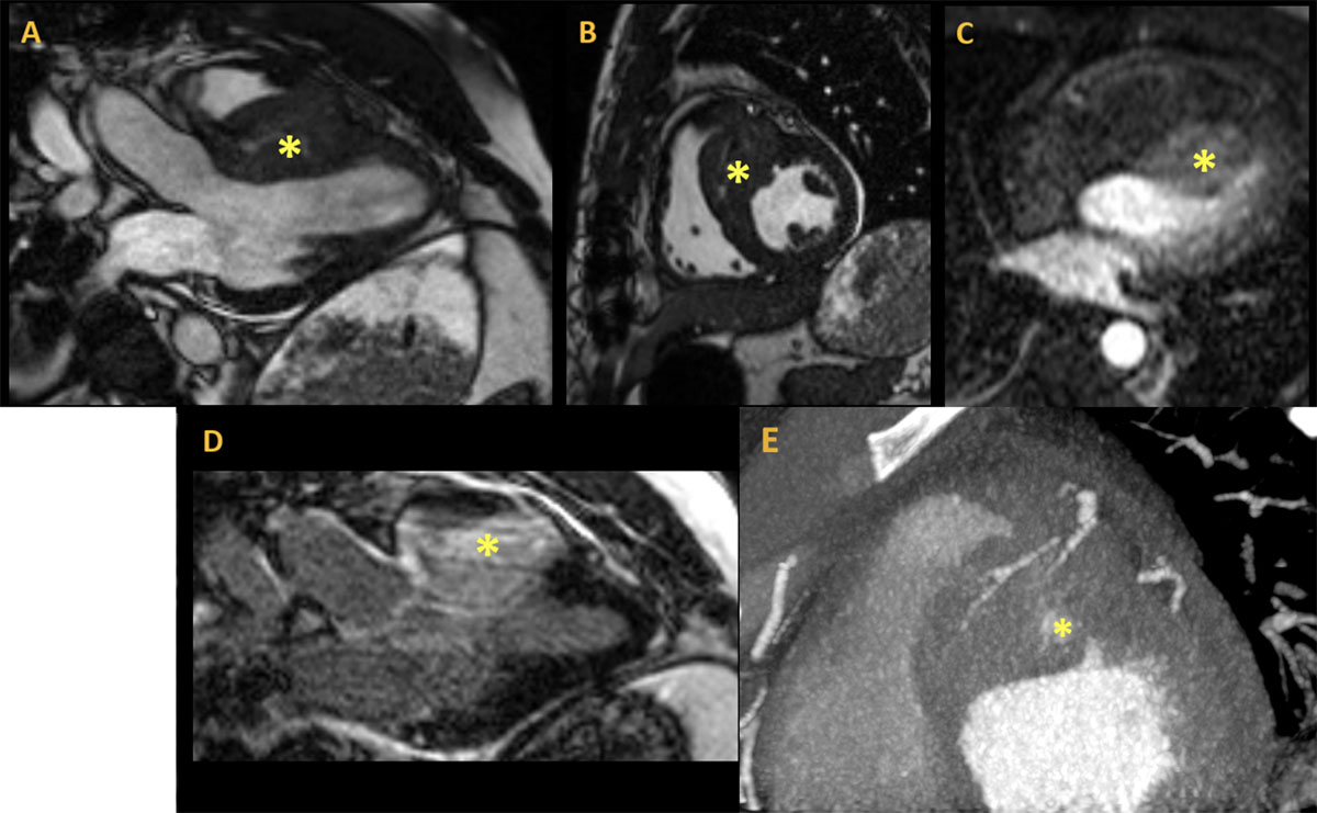
Figure 2
Preoperative cardiac imaging. Magnetic resonance imaging using steady state free precession cine sequence: (A) 3-chamber view, (B) short axis at the mid-level, (C), first pass perfusion, and (D) delayed enhancement after gadolinium showing a large right ventricular (RV) mass measuring 4.9 × 3.7 cm invading the RV free wall, with (E) intense first pass perfusion and significant late gadolinium enhancement cardiac computed tomography demonstrating short axis basal anteroseptal mass protruding into the RV outflow tract with central contrast enhancement and multiple septal perforator feeding vessels.
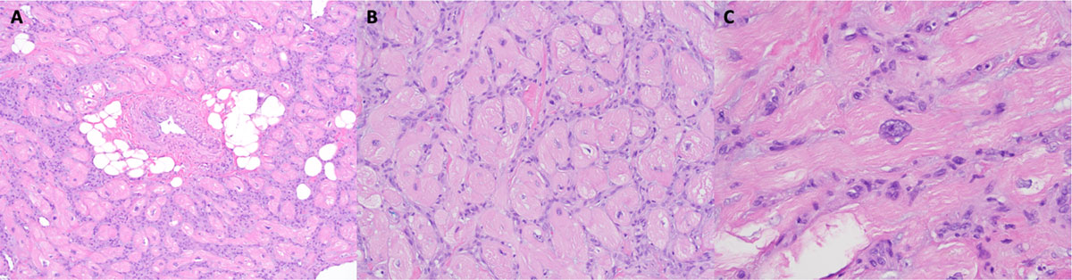
Figure 3
H&E stain at increasing magnification 100×. (A) The resected lesion was composed of disorganized, hypertrophic cardiac myocytes with interstitial fibrosis. (B) There were also scattered thickened intramural arteries, dilated venules, and small collections of adipocytes 200×. (C) At intermediate magnification, the myocytes were haphazardly arranged and enlarged with sarcoplasmic vacuolation and nuclear enlargement 400×. There were scattered enlarged nuclei, some with mild hyperchromasia, irregular nuclear contours, and inconspicuous nucleoli.
Table 1
Cardiac hamartoma literature search. A literature search revealed 30 patients with a diagnosis of hamartoma of mature cardiac myocytes from 1998 to 2020. The hamartoma was localized to the left ventricle in 13 patients, the right atrium in 7 patients, and the right ventricle in 3 patients; 2 patients showcased multiple locations. Ten of these patients (33%) were asymptomatic at time of presentation.1-21 CT: computed tomography; RVR: rapid ventricular response; WPW: Wolff-Parkinson-White.
| YEAR PUBLISHED | AUTHOR | AGE | GENDER | CLINICAL SYMPTOMS | DIAGNOSTIC EVALUATION | TUMOR LOCATION | TUMOR SIZE |
|---|---|---|---|---|---|---|---|
| 1998 | Sturtz et al. | 24 | Male | Hypertension, palpitations, and premature contractions | — | Left ventricle | — |
| 1998 | Burke et al. | 22 | Male | Asymptomatic | Echocardiogram | Right ventricle | 5 cm |
| 1998 | Burke et al. | 28 | Male | WPW syndrome and an episode of syncope | Echocardiogram | Right atrium | — |
| 1998 | Burke et al. | 9 | Male | Sudden death | Autopsy | Right atrium | 1 × 2 cm |
| 2001 | Dinh et al. | 33 | Male | Generalized tachycardia | Echocardiogram | Left ventricle | 4.5 × 3.1 × 4.4 cm |
| 2004 | Chu et al. | 76 | Male | History of hypertension | In surgery | Crista terminalis | 0.5 × 1 × 0.5 cm |
| 2005 | Martínez QM et al. | 33 | Male | Palpitations and dyspnea | Echocardiogram | Left ventricle | 4.5 × 5.5 cm |
| 2008 | Movahedi et al. | 58 | Male | Progressive dyspnea | In surgery | Right atrium | 1.5 × 1 × 0.5 cm |
| 2008 | Fealey et al. | 0.5 | Male | Asymptomatic | — | — | — |
| 2008 | Fealey et al. | 0.5 | Female | Asymptomatic | — | — | — |
| 2008 | Fealey et al. | 1.2 | Male | Asymptomatic | — | — | — |
| 2008 | Fealey et al. | 10 | Male | Asymptomatic | Echocardiogram | Left ventricle | 5 × 3 cm |
| 2008 | Fealey et al. | 16 | Female | Asymptomatic | Echocardiogram | Right ventricle | 8 × 9 cm |
| 2008 | Fealey et al. | 57 | Male | Sudden death | — | — | — |
| 2008 | Fealey et al. | 74 | Male | Exertional dyspnea | — | — | — |
| 2008 | Menon et al. | 16 | Female | Weight loss | Echocardiogram | Right ventricle | 8 × 7 × 3 cm |
| 2008 | Menon et al. | 10 | Male | Asymptomatic | Echocardiogram | Left ventricle | 0.3 × 0.2 cm |
| 2009 | Hsu et al. | 19 | Female | Intermittent palpitations and dizziness | Echocardiogram | Left ventricle | 4 × 7 cm |
| 2009 | Galeone et al. | 56 | Female | Asymptomatic | Chest CT | Pulmonary infundibulum | 9 × 9 × 4 cm |
| 2011 | Dell’Amore et al. | 35 | Female | Palpitations and dyspnea | Echocardiogram | Left ventricle | 4.2 × 3.3 × 2.7 cm |
| 2013 | Raffa et al. | 41 | Female | Chest pain | Echocardiogram | Right atrium | 2.5 × 1.3 cm |
| 2017 | Ayoub et al. | 14 | Male | Asymptomatic | Echocardiogram | Left ventricle | 9 × 5 × 6 cm |
| 2017 | Hadravská et al. | 39 | Female | Ruptured aneurysm and severe pneumonia | Autopsy | Left ventricle | 4.5 × 3 × 3 cm |
| 2017 | Abuzaid et al. | 21 | Female | Chest pain and dyspnea | Echocardiogram | Left ventricle | 1.6 × 1.3 × 1.9 cm |
| 2017 | Liu et al. | 64 | Male | Dyspnea | Echocardiogram | Left ventricle | 2 × 3 cm |
| 2018 | Mantilla-Hernandez et al. | 23 | Female | Paroxysmal nocturnal dyspnea, RVR and edema | Autopsy | Right atrium | — |
| 2018 | Negri et al. | 44 | Female | Asymptomatic | Echocardiogram | Left ventricle | 4 × 3 cm |
| 2018 | Kumari et al. | 43 | Male | Chest pain, orthopnea, and palpitations | Echocardiogram | Left ventricle | 8 × 6 cm |
| 2019 | Fu et al. | 41 | Male | Cough, edema, dyspnea on exertion | Echocardiogram | Right atrium | 2 × 1 × 0.5 cm |
| 2019 | Zhou et al. | 41 | Male | Dyspnea and edema | Echocardiogram | Right atrium | 1.8 × 1.4 cm |